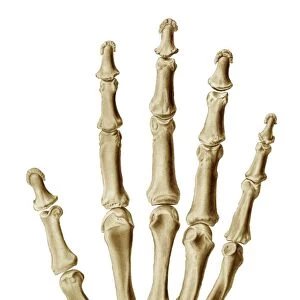
Dorsiflexion of the foot, artwork C016 / 6798

![]()
Wall Art and Photo Gifts from Science Photo Library
Dorsiflexion of the foot, artwork C016 / 6798
Dorsiflexion of the foot. Artwork of the muscles of the foot from the side, with red arrows showing the direction of movement when flexing the foot in the direction of its upper (dorsal) surface (dorsiflexion). The muscles involved are the tibialis anterior, the extensor hallucis longus, the extensor digitorum longus, and the peroneus tertius. These muscles arise in the lower leg, and extend downwards into the foot. The nerve used is the common peroneal nerve. This is the left foot. For the right foot, see C016/6797
Science Photo Library features Science and Medical images including photos and illustrations
Media ID 9243311
© D & L GRAPHICS / SCIENCE PHOTO LIBRARY
Ankle Arthrology Bend Bending Biomechanics Diagram Dorsal Extensor Digitorum Longus Extensor Hallucis Longus Flex Flexing Flexion Foot Joint Lateral Ligament Ligaments Limb Movement Moving Muscles Muscular Physiological Physiology Profile Range Of Movements Tendon Tendons Tibialis Anterior Cutouts Left Foot Musculature
EDITORS COMMENTS
This print titled "Dorsiflexion of the foot, artwork C016 / 6798" showcases the intricate muscles and movements involved in flexing the foot towards its upper surface. Against a clean white background, this detailed illustration highlights the tibialis anterior, extensor hallucis longus, extensor digitorum longus, and peroneus tertius muscles that originate from the lower leg and extend into the foot. The red arrows elegantly guide our attention to the direction of movement when dorsiflexing the foot. This physiological process is made possible by utilizing the common peroneal nerve. While this particular artwork depicts a left foot, an accompanying image (C016/6797) portrays similar details for the right foot. With its emphasis on anatomy and biology, this print provides valuable insights into normal human physiology. It beautifully captures various aspects such as joint flexibility, muscular strength, ligamentous connections, tendons' role in biomechanics – all essential components contributing to healthy movement. As we delve into understanding arthrology and musculature through this visual representation of a lateral view cutout of a human limb's side profile; it becomes evident how crucial these structures are for maintaining optimal mobility. Whether used for educational purposes or personal interest in exploring our body's complexity – this artwork offers an engaging perspective on dorsiflexion and range of movements associated with it.
MADE IN THE USA
Safe Shipping with 30 Day Money Back Guarantee
FREE PERSONALISATION*
We are proud to offer a range of customisation features including Personalised Captions, Color Filters and Picture Zoom Tools
SECURE PAYMENTS
We happily accept a wide range of payment options so you can pay for the things you need in the way that is most convenient for you
* Options may vary by product and licensing agreement. Zoomed Pictures can be adjusted in the Cart.