Light Microscope Collection (#46)

Exploring the Intricacies of Life: A Glimpse through the Light Microscope Unveiling the Wonders of Cerebellum Tissue

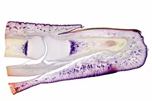
Background imageLight Microscope Collection: Human finger, longitudinal section

Human finger, longitudinal section
Human finger. Light micrograph of a longitudinal section through a finger of a human infant. This shows the bones inside the finger (here, the 1st and 2nd phalanges)

Background imageLight Microscope Collection: Wasp sting, light micrograph

Wasp sting, light micrograph
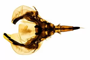
Wasp sting. Light micrograph of the dissected stinging apparatus of a worker wasp (Vespula germanica). In this preparation the three parts of the shaft of the sting have separated into the two

Background imageLight Microscope Collection: Caterpillar skin, light micrograph

Caterpillar skin, light micrograph. This section of part of the skin shows two half segments. The skin is covered in small hairs or setae (black), and brown structures called tubercles and verrucae

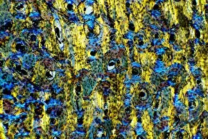
Background imageLight Microscope Collection: Compact bone, light micrograph

Compact bone, light micrograph
Compact bone. Polarised light micrograph of a transverse section through compact bone tissue, showing Haversian canals (circular regions)

Background imageLight Microscope Collection: Dogfish liver area, transverse section

Dogfish liver area, transverse section
Dogfish liver area. Light micrograph of a transverse section through the body of a lesser spotted dogfish (Scyliorhinus canicula), in the region of the liver (lower frame)

Background imageLight Microscope Collection: Bee proboscis, light micrograph

Bee proboscis, light micrograph
Bee proboscis. Polarised light micrograph of the proboscis of the common honey bee (Apis mellifera). The bees proboscis (centre) consists of two outer angled parts (the galea)

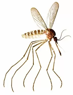
Background imageLight Microscope Collection: Gnat, light micrograph

Gnat, light micrograph
Gnat (Culex pipiens), light micrograph. The gnats body has three major parts: the abdomen, thorax and head. The abdomen is at upper centre

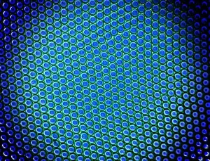
Background imageLight Microscope Collection: Beetle compound eye, light micrograph

Beetle compound eye, light micrograph
Beetle compound eye. Polarised light micrograph of a portion of the compound eye of a great diving beetle (Dytiscus sp.), showing the massed circular lenses of the simple eyes, the ommatidia

Background imageLight Microscope Collection: Kidney, longitudinal section

Kidney, longitudinal section
Kidney. Light micrograph of a longitudinal section through a kidney, showing the outer region (cortex, round the edges) and the inner region (medulla, centre)

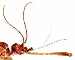
Background imageLight Microscope Collection: Gnat head, light micrograph

Gnat head, light micrograph
Gnat head. Light micrograph of the head (lower left), antennae (upper left), proboscis (lower right) and other mouthparts of a gnat (Culex pipiens)

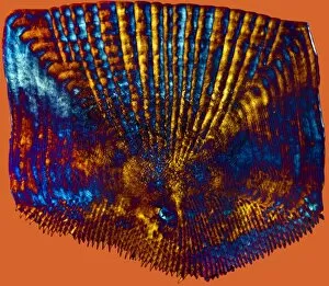
Background imageLight Microscope Collection: Bass scale, light micrograph

Bass scale, light micrograph
Bass scale. Polarised light micrograph of a ctenoid scale from the skin of a bass (Dicentrarchus labrax). The scales are just under the fishs skin and form a protective skeleton

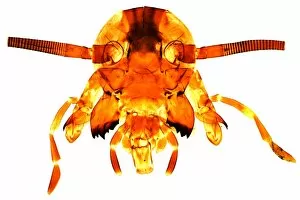
Background imageLight Microscope Collection: Cockroachs head, light micrograph

Cockroachs head, light micrograph
Cockroachs head. Light micrograph of the head and mouthparts of the American cockroach (Periplaneta orientalis). The head consists of two hemispherical compound eyes (top centre)

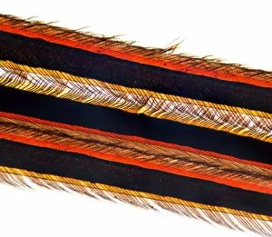
Background imageLight Microscope Collection: Feather vane, light micrograph

Feather vane, light micrograph
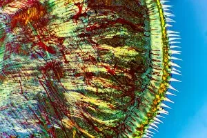
Feather vane. Light micrograph of a part of a vane of a contour feather, from the wing of a bird. Each vane consists of several barbs (black) bearing barbules (thin)

Background imageLight Microscope Collection: Caterpillar proleg, light micrograph

Caterpillar proleg, light micrograph
Caterpillar proleg. Dark-field light micrograph of part of the skin of a caterpillar, showing a proleg (centre). The proleg is a short stubby protrusion

Background imageLight Microscope Collection: Spider spinnerets, light micrograph

Spider spinnerets, light micrograph
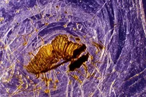
Spider spinnerets. Light micrograph of the end of the abdomen of a spider, showing the six spinnerets. Two spinnerets are on the top of two flattened cones (right)

Background imageLight Microscope Collection: Eel scale, light micrograph

Eel scale, light micrograph
Eel scale. Polarised light micrograph of a scale from the skin of an eel (Anguilla anguilla). The scales are just under the skin and form a protective skeleton

Background imageLight Microscope Collection: Dogfish head, transverse section

Dogfish head, transverse section
Dogfish head. Light micrograph of a transverse section through the head of a lesser spotted dogfish (Scyliorhinus canicula), in the region of the eyes (upper left and upper right)

Background imageLight Microscope Collection: Female flea, light micrograph

Female flea, light micrograph
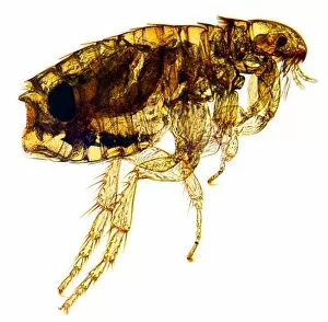
Female flea. Polarised light micrograph of the whole body of a female Pulex flea. The head of the flea is at upper right, with a simple eye (ocellus), two antennae, and piercing, sucking mouthparts

Background imageLight Microscope Collection: Intervertebral disc, light micrograph

Intervertebral disc, light micrograph
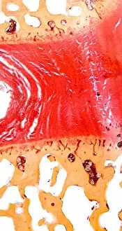
Intervertebral disc. Light micrograph of a transverse section through an intervertebral disc (red) and the surrounding tissue (brown)

Background imageLight Microscope Collection: Heel skin tissue, light micrograph

Heel skin tissue, light micrograph
Heel skin tissue. Polarised light micrograph of a transverse section through skin from the heel of a human foot. The sole of the foot has to withstand the weight of the body

Background imageLight Microscope Collection: Lamprey gill region, light micrograph

Lamprey gill region, light micrograph
Lamprey gill. Light micrograph of a transverse section through the gill region of a lamprey (Petromyzon marinus), a parasitic fish. The spinal area is at upper centre

Background imageLight Microscope Collection: Frog bladder tissue, light micrograph

Frog bladder tissue, light micrograph
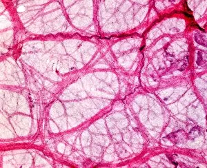
Frog bladder tissue. Light micrograph of tissue from the bladder of a frog (Rana temperaria), showing the smooth muscle network (pink)

Background imageLight Microscope Collection: Breast tissue, light micrograph

Breast tissue, light micrograph
Breast tissue. Light micrograph of a section through tissue from the human mammary gland during pregnancy. The breast is normally made up of fibro-fatty connective tissue

Background imageLight Microscope Collection: Caterpillar spiracle, light micrograph

Caterpillar spiracle, light micrograph
Caterpillar spiracle. Polarised light micrograph of part of the skin of a caterpillar, showing an air hole (spiracle, dark oval, lower right) leading to a breathing tube (trachea)

Background imageLight Microscope Collection: Rabbit eye, longitudinal section

Rabbit eye, longitudinal section
Rabbit eye. Light micrograph of a longitudinal section through the eye of a rabbit (Oryctolagus cuniculus). The rabbit has a typical mammalian eye structure

Background imageLight Microscope Collection: Backswimmers head, light micrograph

Backswimmers head, light micrograph
Backswimmers head. Light micrograph of the head and mouthparts of the backswimmer (Notonecta sp.), also known as the greater water boatman

Background imageLight Microscope Collection: Shark skin, light micrograph

Shark skin, light micrograph
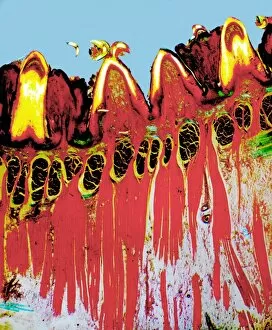
Shark skin. Light micrograph of a section through the skin of a shark, showing two dermal denticles. These bony structures cover the whole skin of the shark
Background imageLight Microscope Collection: Taste buds, light micrograph

Taste buds, light micrograph
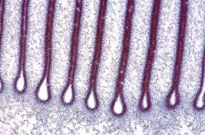
Taste buds. Coloured light micrograph of a section through the tongue, showing taste buds (round, purple). The taste buds are within papillae (projections) located on the surface of the tongue

Background imageLight Microscope Collection: Stomach pylorus glands, light micrograph

Stomach pylorus glands, light micrograph
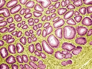
Stomach pylorus glands. Light micrograph of a section through glands (pink) in the pylorus region of the stomach. Pyloric glands contain mucus cells and G cells that secrete gastrin

Background imageLight Microscope Collection: Sheeps hoof, light micrograph

Sheeps hoof, light micrograph
Sheeps hoof. Polarised light micrograph of a transverse section through the hoof of a sheep. In sheep and other hooved animals, the hoof is made of the same material as nails and claws

Background imageLight Microscope Collection: Pneumonia infection, light micrograph

Pneumonia infection, light micrograph
Pneumonia infection. Light micrograph of a section through lung tissue from a patient with bronchial pneumonia. Shown here is the blockage and breakdown of the air sacs

Background imageLight Microscope Collection: Fern spore cases, light micrograph

Fern spore cases, light micrograph
Fern spore cases. Polarised light micrograph of a frond of the maidenhair fern (Adiantum capillus- veneris), showing the sporangia (black, centre left) which contain the ferns spores
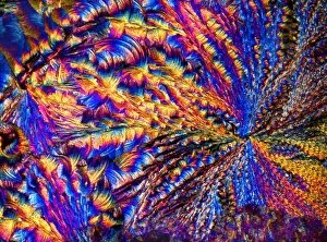
Background imageLight Microscope Collection: Lichen, light micrograph

Lichen, light micrograph
Lichen. Light micrograph of a section through tissue of a lichen. Lichens are symbiotic associations of a fungus (the mycobiont) with a photosynthetic partner (the photobiont)

Background imageLight Microscope Collection: Mushroom, light micrograph

Mushroom, light micrograph
Mushroom. Light micrograph (LM) of a section through a mushroom, Agaricus sp. (formerly Psalliota sp.), showing its stalk (orange) and gills (long white projections)

Background imageLight Microscope Collection: Mushroom gills, light micrograph

Mushroom gills, light micrograph
Mushroom gills. Light micrograph (LM) of a section through the gills of a mushroom, Agaricus sp. (formerly Psalliota sp.)

Background imageLight Microscope Collection: Silver nitrate crystals, LM

Silver nitrate crystals, LM
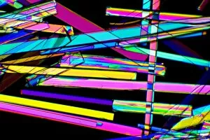
Silver nitrate crystals. Polarised light micrograph (LM) of silver nitrate (AgNO3) crystals. Silver nitrate is used in photography and as a test for chloride, bromide and iodide ions

Background imageLight Microscope Collection: Magnesium chloride crystals, LM

Magnesium chloride crystals, LM
Magnesium chloride. Polarised light micrograph (LM) of crystals of magnesium chloride (MgCl2). Typically, magnesium chloride forms complexes with other molecules; the hygroscopic hexahydrate

Background imageLight Microscope Collection: Magnesium sulphate crystals, LM

Magnesium sulphate crystals, LM
Magnesium sulphate. Polarised light micrograph of crystals of magnesium sulphate (MgSO4), a chemical used medicinally and in the textile industry

Background imageLight Microscope Collection: Spinal cord, transverse section

Spinal cord, transverse section
Spinal cord. Light micrograph of a transverse section through a human spinal cord. The spinal cord has a central canal around which are the dorsal and ventral horns (orange)

Background imageLight Microscope Collection: Dog tongue tissue, light micrograph

Dog tongue tissue, light micrograph
Dog tongue tissue. Light micrograph of a transverse section through the tongue of a dog, showing the tongues surface (across top)

Background imageLight Microscope Collection: Foot skin tissue, light micrograph

Foot skin tissue, light micrograph
Foot skin tissue. Light micrograph of a transverse section through skin from a human foot. The skin is made up of an outer epidermis (across top)

Background imageLight Microscope Collection: Nerve cell trauma response

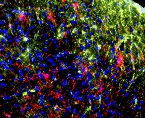
Nerve cell trauma response. Fluorescent light micrograph of a section through a spinal cord affected by multiple sclerosis (MS)

Background imageLight Microscope Collection: Brain tissue, light micrograph

Brain tissue, light micrograph
Brain tissue. Light micrograph of a section through tissue from the mammalian brain, in this case from the cerebellum. The cerebellum is the part of the brain that controls sensory perception

Background imageLight Microscope Collection: Skin sweat glands, light micrograph

Skin sweat glands, light micrograph
Skin sweat glands. Light micrograph of a transverse section through skin from a human foot. The skin is made up of an outer epidermis (across top) and a lower epidermis (deep purple)

Background imageLight Microscope Collection: Cat tongue tissue, light micrograph

Cat tongue tissue, light micrograph
Cat tongue tissue. Polarised light micrograph of a transverse section through a cats tongue. The outer stratified epithelium (right) has spike-like (white) keratinised papillae

Background imageLight Microscope Collection: Tongue tissue, light micrograph

Tongue tissue, light micrograph
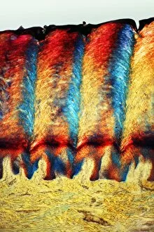
Tongue tissue. Polarised light micrograph of a transverse section through a tongue, showing the surface (across top). Here, there are four rounded fungiform papillae

Background imageLight Microscope Collection: Zeolite crystals, light micrograph

Zeolite crystals, light micrograph
Zeolite crystals. Polarised light micrograph of crystals of zeolite from the Giants Causeway, Northern Ireland. Zeolites are hydrated aluminosilicate minerals and have a micro-porous structure

Background imageLight Microscope Collection: Light Micrograph (LM): plant cell chloroplasts - the site where photosynthesis takes place

Light Micrograph (LM): plant cell chloroplasts - the site where photosynthesis takes place
LRDS-192 Light Micrograph (LM): plant cell chloroplasts - the site where photosynthesis takes place David Spears (Last Refuge)


For sale as Licensed Images

Choose your image, Select your licence and Download the media

Exploring the Intricacies of Life: A Glimpse through the Light Microscope Unveiling the Wonders of Cerebellum Tissue: Witness the intricate network of cells in cerebellum tissue as seen through a captivating light micrograph. Nerve and Glial Cells Revealed: Dive into the mesmerizing world of nerve and glial cells, captured in stunning detail by a powerful light microscope. Unraveling Chemical Structures: Behold the ethereal beauty of copper and magnesium sulphate crystals, magnified under a light micrograph to expose their delicate arrangements. The Hidden Beauty Within Caffeine Crystals: Marvel at the crystalline structures of caffeine, brought to life through an enchanting light micrograph that unveils its hidden allure. Journey into Hippocampus Brain Tissue: Embark on an extraordinary expedition deep within hippocampus brain tissue, where mysteries unfold with every glance through our remarkable light microscope lens. Illuminating Glial Cells' Secrets: Delve into the enigmatic realm of glial cells as they reveal their intricate patterns and connections in a mesmerizing confocal light micrograph. HeLa Cells Exposed - A Window to Immortality? Explore HeLa cells like never before with our cutting-edge technology, providing unprecedented insight into these immortalized wonders under brightfield illumination (C017 / 8299). Peering Into Purkinje Nerve Cells in Cerebellum: Discover the elegance and complexity displayed by Purkinje nerve cells residing within cerebellar tissue when observed using a high-resolution light microscope. Dicotyledon Plant Stem's Inner Workings Revealed. Get lost amidst nature's marvels as we uncover the intricacies present within dicotyledon plant stems using our advanced techniques for capturing breathtakingly detailed images under brightfield illumination. Unveiling the Secrets of Kidney Tubules.