
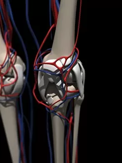
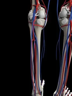
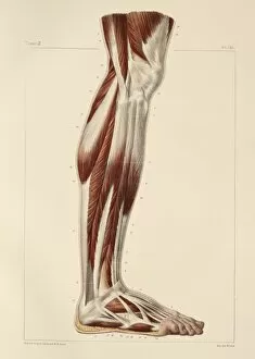
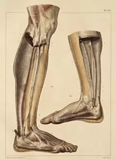
Lower Leg Collection
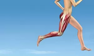
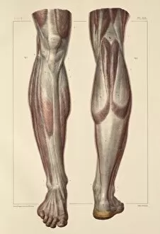
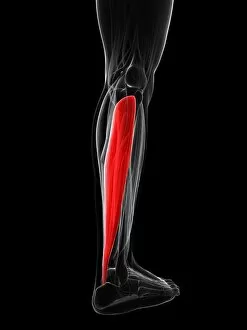
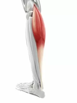
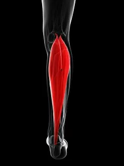
"Exploring the Lower Leg: From Muscles to Artwork" Leg muscles in running


For sale as Licensed Images
Choose your image, Select your licence and Download the media
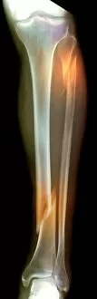
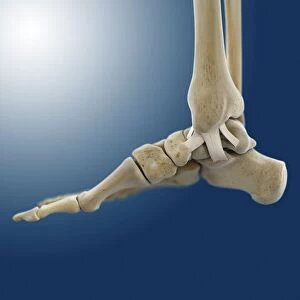
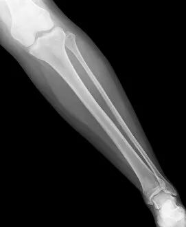
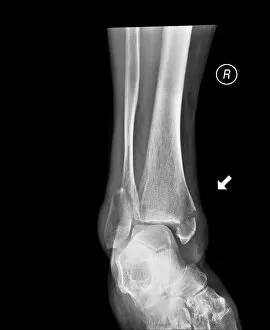
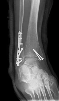
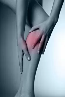
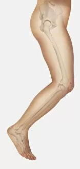
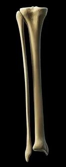
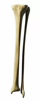
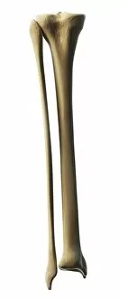
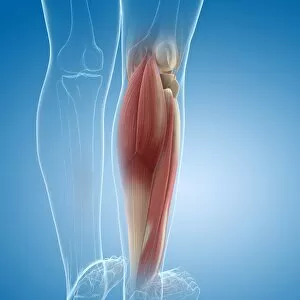
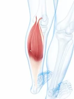
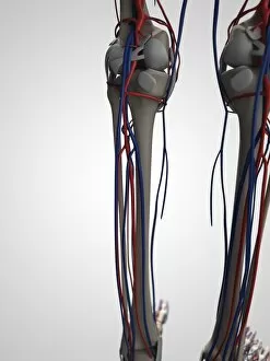
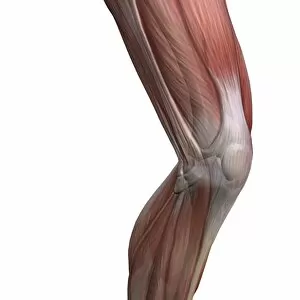
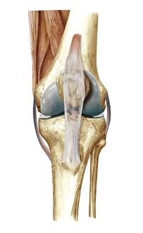
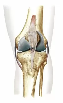
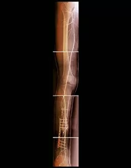
"Exploring the Lower Leg: From Muscles to Artwork" Leg muscles in running: Discover the intricate network of leg muscles that power your every stride on the track or trail. Unveiling an Artistic Perspective: Dive into captivating artwork depicting the beauty and strength muscles in motion. A Closer Look at Fractures: Witness a double fracture through an X-ray, highlighting the resilience and healing process of the lower leg. Inner Ankle Ligaments Revealed: Delicate yet crucial, admire artwork showcasing inner ankle ligaments that provide stability during movement. Peering Inside with X-rays: Observe a normal lower leg through an X-ray, appreciating its complex structure and functionality. The Impact of a Broken Ankle: Gain insight into the severity of a broken ankle as captured by an X-ray, emphasizing the importance of proper care and treatment. Triumph Over Adversity: Marvel at an amputated lower leg depicted in an X-ray, symbolizing resilience and determination in overcoming challenges. Restoring Stability with Pins: Explore how medical intervention can restore stability to a broken ankle through a pinned X-ray image. Addressing Lower Leg Pain: Learn about common causes and potential remedies for discomfort experienced within this vital part of our body's foundation. Saul's Legs Study Series Painting: Immerse yourself in Saul's artistic masterpiece series capturing various aspects of human legs' gracefulness and strength. Invention Drawing Unveiled - Sketching Lower Leg Bones: Witness creativity come alive as inventors sketch out ideas for improving understanding or treating lower leg conditions. Jumping Into Action - Diagram Showing Bones Inside Human Leg Ready to Jump : Visualize bone structures inside your legs preparing for action—ready to propel you forward with each leap. 13. Seated Comfortably - Diagram Showing Bones Inside Human Leg in a Seated Position.