
Lumbar Collection
"Lumbar: Exploring the Intricacies of the Human Spine and Abdomen" Delving into the depths of our body's core


For sale as Licensed Images
Choose your image, Select your licence and Download the media
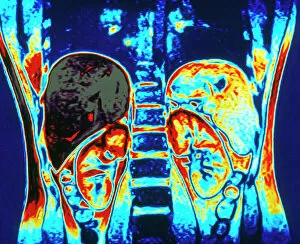
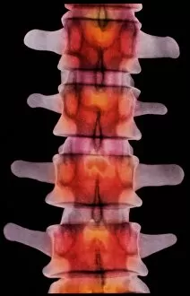
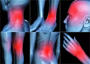
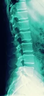
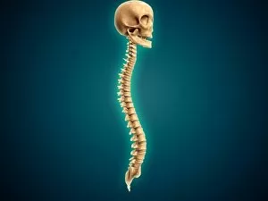
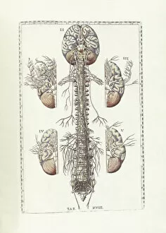
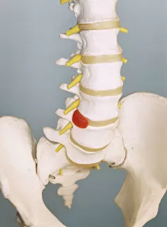
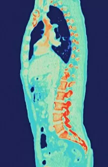
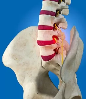
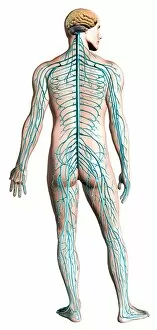
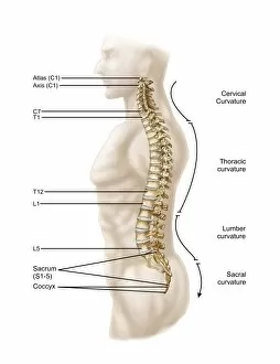
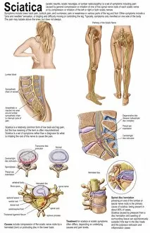
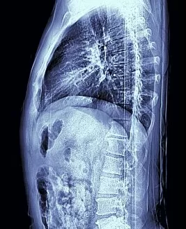
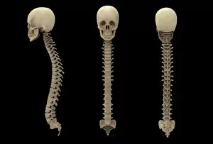
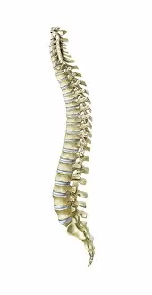
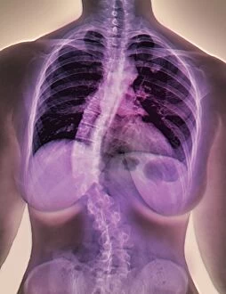
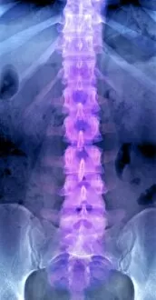
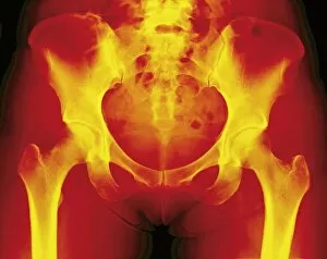
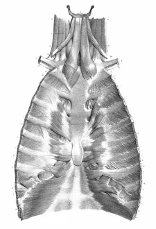
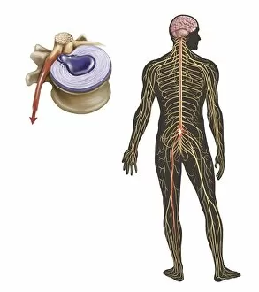
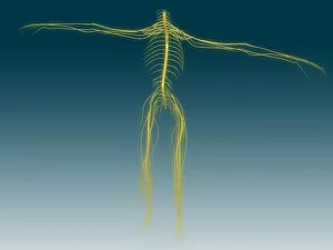
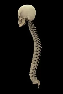
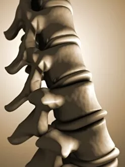
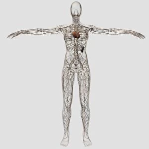
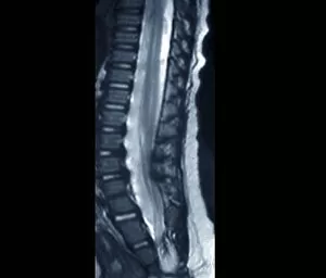
"Lumbar: Exploring the Intricacies of the Human Spine and Abdomen" Delving into the depths of our body's core, a colour MRI scan reveals the intricate beauty of kidneys and liver in vivid detail. Body pain can be debilitating, but through art, we find solace in expressing our struggles and seeking healing for our lumbar region. A coloured X-ray unveils the delicate architecture vertebrae, providing a glimpse into the foundation that supports our spine's strength and flexibility. Behold a normal spine captured by an X-ray - a testament to its resilience and ability to withstand life's daily challenges. In this conceptual image, a human skull stands as a reminder of how intricately connected it is to our spinal cord, emphasizing their vital role in maintaining bodily functions. Bartholomeo Eustachi's masterpiece "The Science of Human Anatomy" takes us on an enlightening journey through centuries-old knowledge about lumbar anatomy. The excruciating pain caused by a slipped disc reminds us of the fragility within our lower back region - urging us to prioritize its care and seek proper treatment when needed. Through an MRI scan capturing a normal torso, we witness firsthand how crucial it is to maintain good lumbar health for overall well-being. Artwork depicting a slipped intervertebral disc serves as both an eye-opening visual representation and an empathetic portrayal of those grappling with this common yet painful condition. An X-ray showcasing a healthy lower back reinforces the importance of preventive measures such as exercise, proper posture, and regular check-ups to preserve optimal spinal alignment throughout life. Pinned vertebrae immobilized by medical intervention remind us that sometimes support from external sources becomes necessary for restoring stability within our lumbar region