Macromolecule Collection (#10)

Macromolecules, the building blocks of life, are at the forefront of scientific innovation

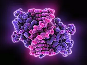
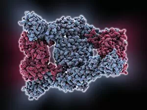
Background imageMacromolecule Collection: Haemoglobin S, molecular model

Haemoglobin S, molecular model
Haemoglobin S. Molecular model of the mutant form of haemoglobin (haemoglobin S) that causes sickle cell anaemia. This is deoxyhaemoglobin S, the molecule in its non-oxygen bound state

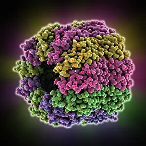
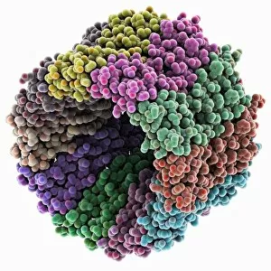
Background imageMacromolecule Collection: Alpha 3 bacteriophage capsid

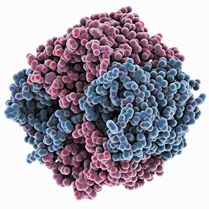
Alpha 3 bacteriophage capsid, molecular model. Bacteriophages are viruses that infect bacteria, with the capsid forming the bacteriophage head. A capsid has subunits called capsomeres

Background imageMacromolecule Collection: Opsin molecule C016 / 0635

Opsin molecule C016 / 0635
Opsin. Molecular model of a ligand-free opsin molecule. Opsins are found in photoreceptor cells (rods and cones) in the retina of the eye
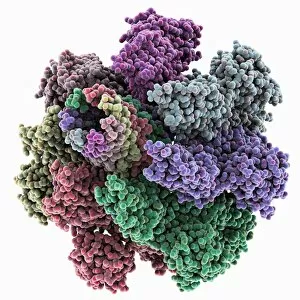
Background imageMacromolecule Collection: Iron-regulatory protein bound to RNA C015 / 6691

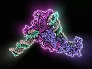
Iron-regulatory protein bound to RNA C015 / 6691
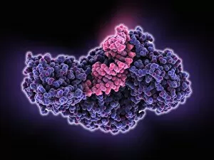
Iron-regulatory protein bound to RNA, molecular model. Iron regulatory protein 1 (IRP1, purple) bound to a short strand of RNA (ribonucleic acid, pink) that includes iron-responsive elements (IREs)
Background imageMacromolecule Collection: Iron-regulatory protein bound to RNA C015 / 6690

Iron-regulatory protein bound to RNA C015 / 6690
Iron-regulatory protein bound to RNA, molecular model. Iron regulatory protein 1 (IRP1, blue) bound to a short strand of RNA (ribonucleic acid, pink) that includes iron-responsive elements (IREs)

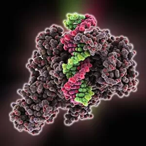
Background imageMacromolecule Collection: DNA hybrid duplex, molecular model

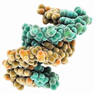
DNA hybrid duplex, molecular model. This model shows a chimeric junction, where a DNA (deoxyribonucleic acid) strand changes from one form to another

Background imageMacromolecule Collection: RSV virus fusion glycoprotein C015 / 6689

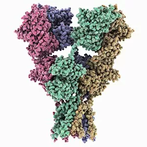
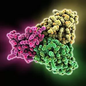
RSV virus fusion glycoprotein C015 / 6689
RSV virus fusion glycoprotein, molecular model. This is a fusion glycoprotein of human respiratory syncytial virus (RSV). This glycoprotein is used by the virus to invade host cells

Background imageMacromolecule Collection: RSV virus fusion glycoprotein C015 / 6688

RSV virus fusion glycoprotein C015 / 6688
RSV virus fusion glycoprotein, molecular model. This is a fusion glycoprotein of human respiratory syncytial virus (RSV). This glycoprotein is used by the virus to invade host cells

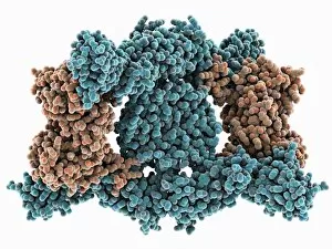
Background imageMacromolecule Collection: Adenovirus penton base protein

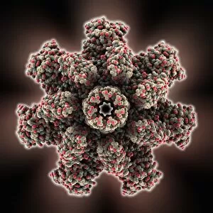
Adenovirus penton base protein, molecular model. This protein molecule is a subunit called a penton, forming the vertices of the capsid of this adenovirus

Background imageMacromolecule Collection: Aquaporin membrane protein C015 / 5922

Aquaporin membrane protein C015 / 5922
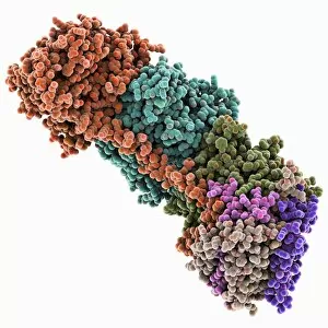
Aquaporin membrane protein, molecular model. Aquaporins are membrane proteins that form channels (lower right) that help water molecules pass in and out of cells

Background imageMacromolecule Collection: Kinesin motor protein dimer C015 / 5921

Kinesin motor protein dimer C015 / 5921
Kinesin motor protein dimer, molecular model. Kinesin is a motor protein that moves along microtubule filaments in cells. It does so by forming a dimer, the heads of which walk along the microtubule

Background imageMacromolecule Collection: Kinesin motor protein dimer C015 / 5920

Kinesin motor protein dimer C015 / 5920
Kinesin motor protein dimer, molecular model. Kinesin is a motor protein that moves along microtubule filaments in cells. It does so by forming a dimer, the heads of which walk along the microtubule

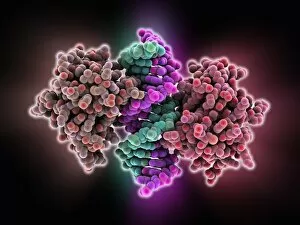
Background imageMacromolecule Collection: Transcription repressor protein and DNA C015 / 5810

Transcription repressor protein and DNA C015 / 5810
Transcription repressor protein and DNA, molecular model. The repressor protein (green) is binding to a strand of DNA (deoxyribonucleic acid, pink and purple)

Background imageMacromolecule Collection: Chaperonin folding protein C015 / 5560

Chaperonin folding protein C015 / 5560
Chaperonin folding protein, molecular model. Chaperonins are proteins that provide favourable conditions for the correct folding of other proteins

Background imageMacromolecule Collection: Chaperonin folding protein C015 / 5559

Chaperonin folding protein C015 / 5559
Chaperonin folding protein, molecular model. Chaperonins are proteins that provide favourable conditions for the correct folding of other proteins

Background imageMacromolecule Collection: Heat shock transcription factor and DNA C015 / 5558

Heat shock transcription factor and DNA C015 / 5558
Heat shock transcription factor and DNA. Molecular model of the binding domain region (purple) of a heat shock protein transcription factor bound to DNA (pink, deoxyribonucleic acid)

Background imageMacromolecule Collection: Oxidoreductase enzyme complex C015 / 6554

Oxidoreductase enzyme complex C015 / 6554
Oxidoreductase enzyme complex, molecular model. This is the membrane-bound domain formed from of a complex of NADH-quinone oxidoreductase subunits

Background imageMacromolecule Collection: Oxidoreductase enzyme complex C015 / 6553

Oxidoreductase enzyme complex C015 / 6553
Oxidoreductase enzyme complex, molecular model. This is the membrane-bound domain formed from of a complex of NADH-quinone oxidoreductase subunits

Background imageMacromolecule Collection: SMAD4 protein domain bound to DNA C015 / 6552

SMAD4 protein domain bound to DNA C015 / 6552
SMAD4 protein domain bound to DNA, molecular model. This strand of DNA (deoxyribonucleic acid, green and pink) is surrounded by MH1 domains of the SMAD4 (Mothers against decapentaplegic homolog 4)

Background imageMacromolecule Collection: SMAD4 protein domain bound to DNA C015 / 6551

SMAD4 protein domain bound to DNA C015 / 6551
SMAD4 protein domain bound to DNA, molecular model. This strand of DNA (deoxyribonucleic acid, red and blue) is surrounded by MH1 domains of the SMAD4 (Mothers against decapentaplegic homolog 4)

Background imageMacromolecule Collection: Vitamin B12 transport protein C015 / 5824

Vitamin B12 transport protein C015 / 5824
Vitamin B12 transport protein, molecular model. This transmembrane protein, known as BTUB, is from the Escherichia coli bacterium

Background imageMacromolecule Collection: Vitamin B12 transport protein C015 / 5823

Vitamin B12 transport protein C015 / 5823
Vitamin B12 transport protein, molecular model. This transmembrane protein, known as BTUB, is from the Escherichia coli bacterium

Background imageMacromolecule Collection: Ionotropic glutamate receptor C015 / 5813

Ionotropic glutamate receptor C015 / 5813
Ionotropic glutamate receptor, molecular model. When glutamate binds to this receptor, it opens up trans-membrane ion channels vital for the functioning of the nervous system

Background imageMacromolecule Collection: Ionotropic glutamate receptor C015 / 5812

Ionotropic glutamate receptor C015 / 5812
Ionotropic glutamate receptor, molecular model. When glutamate binds to this receptor, it opens up trans-membrane ion channels vital for the functioning of the nervous system

Background imageMacromolecule Collection: Transcription repressor protein and DNA C015 / 5811

Transcription repressor protein and DNA C015 / 5811
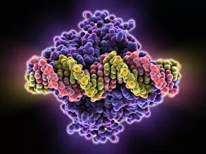
Transcription repressor protein and DNA, molecular model. The repressor protein (purple) is binding to a strand of DNA (deoxyribonucleic acid, yellow and pink)

Background imageMacromolecule Collection: Apoptosome cell death protein C015 / 6109

Apoptosome cell death protein C015 / 6109
Apoptosome cell death protein, molecular model. Apoptosomes are large protein structures formed during programmed cell death (PCD, also called apoptosis)

Background imageMacromolecule Collection: Beta secretase enzyme, molecular model C015 / 5277

Beta secretase enzyme, molecular model C015 / 5277
Beta secretase enzyme. Molecular model of the enzyme beta secretase bound to an inhibitor molecule. Beta secretase is a membrane-associated aspartic protease

Background imageMacromolecule Collection: Self-assembled DNA triangle C015 / 5423

Self-assembled DNA triangle C015 / 5423
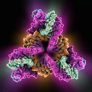
Self-assembled DNA triangle. Molecular model of DNA (deoxyribonucleic acid) strands forming what is called a tensegrity triangle

Background imageMacromolecule Collection: Self-assembled DNA triangle C015 / 5422

Self-assembled DNA triangle C015 / 5422
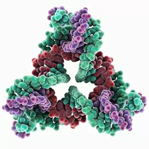
Self-assembled DNA triangle. Molecular model of DNA (deoxyribonucleic acid) strands forming what is called a tensegrity triangle

Background imageMacromolecule Collection: Interferon antagonism by viral protein C015 / 5421

Interferon antagonism by viral protein C015 / 5421
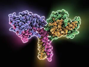
Interferon (IFN) antagonism by viral protein. Molecular model of an orthopoxvirus IFN-gamma-binding protein tetramer bound to an IFN-gamma dimer

Background imageMacromolecule Collection: Interferon antagonism by viral protein C015 / 5420

Interferon antagonism by viral protein C015 / 5420
Interferon (IFN) antagonism by viral protein. Molecular model of an orthopoxvirus IFN-gamma-binding protein tetramer bound to an IFN-gamma dimer

Background imageMacromolecule Collection: Caspase 6 enzyme C015 / 5700

Caspase 6 enzyme C015 / 5700
Capase 6 enzyme, molecular model. Caspases are proteases, enzymes that cleave proteins and play a role in apoptosis (programmed cell death)

Background imageMacromolecule Collection: Chaperonin folding protein C015 / 5697

Chaperonin folding protein C015 / 5697
Chaperonin folding protein, molecular model. Chaperonins are proteins that provide favourable conditions for the correct folding of other proteins

Background imageMacromolecule Collection: Chaperonin folding protein C015 / 5698

Chaperonin folding protein C015 / 5698
Chaperonin folding protein, molecular model. Chaperonins are proteins that provide favourable conditions for the correct folding of other proteins

Background imageMacromolecule Collection: Caspase 6 enzyme C015 / 5699

Caspase 6 enzyme C015 / 5699
Capase 6 enzyme, molecular model. Caspases are proteases, enzymes that cleave proteins and play a role in apoptosis (programmed cell death)

Background imageMacromolecule Collection: Influenza virus protein domain C015 / 6231

Influenza virus protein domain C015 / 6231
Influenza virus protein domain, molecular model. This is an effector domain, a non-structural protein fragment that facilitates interactions

Background imageMacromolecule Collection: Cholera cytolysin cell toxin C015 / 6228

Cholera cytolysin cell toxin C015 / 6228
Cholera cytolysin cell toxin, molecular model. This is the Vibrio cholerae cytolysin (VCC) toxin from the bacterium that causes cholera

Background imageMacromolecule Collection: Cholera cytolysin cell toxin C015 / 6229

Cholera cytolysin cell toxin C015 / 6229
Cholera cytolysin cell toxin, molecular model. This is the Vibrio cholerae cytolysin (VCC) toxin from the bacterium that causes cholera

Background imageMacromolecule Collection: Influenza virus protein domain C015 / 6230

Influenza virus protein domain C015 / 6230
Influenza virus protein domain, molecular model. This is an effector domain, a non-structural protein fragment that facilitates interactions

Background imageMacromolecule Collection: Sodium V-ATPase rotor ring C015 / 5379

Sodium V-ATPase rotor ring C015 / 5379
Sodium V-ATPase rotor ring, molecular model. V-ATPases (vacuolar-type ATPase) are evolutionarily ancient enzymes that helps transport ions across cell membranes

Background imageMacromolecule Collection: Methyltransferase complexed with DNA C015 / 6425

Methyltransferase complexed with DNA C015 / 6425
Methyltransferase complexed with DNA, molecular model. The strand of DNA (deoxyribonucleic acid, red and green) is enclosed by DNA methyltransferase 1 (DNMT-1, brown)

Background imageMacromolecule Collection: Methyltransferase complexed with DNA C015 / 6424

Methyltransferase complexed with DNA C015 / 6424
Methyltransferase complexed with DNA, molecular model. The strand of DNA (deoxyribonucleic acid, red and pink) is enclosed by DNA methyltransferase 1 (DNMT-1, blue)

Background imageMacromolecule Collection: Sodium V-ATPase rotor ring C015 / 5378

Sodium V-ATPase rotor ring C015 / 5378
Sodium V-ATPase rotor ring, molecular model. V-ATPases (vacuolar-type ATPase) are evolutionarily ancient enzymes that helps transport ions across cell membranes

Background imageMacromolecule Collection: VSIV virus protein complex C015 / 6423

VSIV virus protein complex C015 / 6423
VSIV virus protein complex, molecular model. This decameric (10-part) circular structure is a complex of nucleoproteins (nucleocapsid protein)

Background imageMacromolecule Collection: Scorpion toxin bound to antibody C015 / 5158

Scorpion toxin bound to antibody C015 / 5158
Scorpion toxin bound to antibody, molecular model. The toxin is Cn2 from a Centruroides noxius scorpion. The antibodies are human single-chain antibody fragments (scFv) called 9004G

Background imageMacromolecule Collection: VSIV virus protein complex C015 / 6422

VSIV virus protein complex C015 / 6422
VSIV virus protein complex, molecular model. This decameric (10-part) circular structure is a complex of nucleoproteins (nucleocapsid protein)

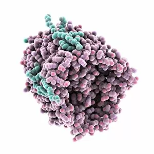
Background imageMacromolecule Collection: RuBisCO activase enzyme

RuBisCO activase enzyme, molecular model. This is RuBisCO activase (Rca) from the tobacco plant (Nicotiana tabacum). RuBisCO stands for ribulose-1, 5-bisphosphate carboxylase oxygenase

Background imageMacromolecule Collection: Chromatin remodelling factor and DNA C015 / 5156

Chromatin remodelling factor and DNA C015 / 5156
Chromatin remodelling factor and DNA, molecular model. The strands of DNA (deoxyribonucleic acid) are at left and right (both red and green). This chromatin remodelling factor (purple) is ISW1a


For sale as Licensed Images

Choose your image, Select your licence and Download the media

Macromolecules, the building blocks of life, are at the forefront of scientific innovation. Nanotube technology has revolutionized various fields, enabling advancements in medicine and electronics. In this captivating computer artwork, we witness the intricate Zinc fingers binding to a DNA strand, showcasing their crucial role in gene regulation. Carbon nanotubes have also emerged as remarkable materials with immense potential. Their unique structure and properties make them ideal for applications ranging from energy storage to drug delivery systems. Computer-generated images depict these carbon nanotubes in all their glory. The SARS coronavirus protein is another macromolecule that has garnered significant attention due to its role in viral infection. Scientists tirelessly study it to develop effective treatments against deadly outbreaks. Computer models allow us to explore complex structures like Bacteriophage phi29—a virus that infects bacteria—providing insights into its mechanisms and aiding in the development of targeted therapies. Simian immunodeficiency virus (SIV), closely related to HIV, poses a global health challenge. Understanding its macromolecular components helps researchers devise strategies for prevention and treatment. Rhodopsin protein molecule captures our imagination with its vital function in vision. Its elegant structure enables light detection and initiates visual signals within our eyes. TFAM transcription factor bound to DNA C015/7059 showcases how macromolecules regulate gene expression by interacting with specific regions on DNA strands—an essential process for cell functioning and development. These glimpses into the world of macromolecules highlight their significance across diverse disciplines—from cutting-edge technologies like nanotube engineering to unraveling infectious diseases or understanding fundamental biological processes. As scientists continue exploring these fascinating molecules, they pave the way for groundbreaking discoveries that shape our future.