Magnified Image Collection (#10)

"Exploring the unseen wonders of the microscopic world: from particle tracks to intricate tissues and beyond

Background imageMagnified Image Collection: Oestradiol, female sex hormone

Oestradiol, female sex hormone
Female sex hormone. Polarised light micrograph of crystals of Oestradiol, also known as beta- Estradiol. This is the most potent of the six naturally-occurring oestrogens

Background imageMagnified Image Collection: Serotonin crystals, light micrograph

Serotonin crystals, light micrograph
Serotonin. Polarised light micrograph of serotonin, a neurotransmitter. Serotonin is derived from tryptophan, and is found in the vertebrate brain

Background imageMagnified Image Collection: Balancing stone from inner ear, SEM

Balancing stone from inner ear, SEM
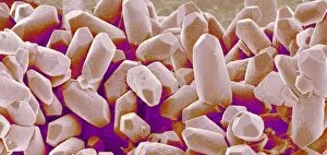
Balancing stone from inner ear. Coloured scanning electron micrograph (SEM) of crystals of calcium carbonate on the surface of a human otolith

Background imageMagnified Image Collection: Syenite, thin section, polarized LM

Syenite, thin section, polarized LM
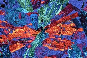
Polarised light micrograph of a thin section of syenite, a coarse-grained igneous rock, containing the mineral augite. Augite occurs as a prismatic crystal which in cross-section appears almost

Background imageMagnified Image Collection: Mica schist, thin section, polarised LM

Mica schist, thin section, polarised LM
Polarised light micrograph of a thin section of mica schist, a type of metamorphic rock. Magnification: x12 at 35mm size

Background imageMagnified Image Collection: Limestone, thin section, polarised LM

Limestone, thin section, polarised LM
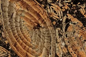
Nummulitic limestone. Polarised light micrograph of a thin section of a Nummulitic limestone. The large coloured objects are the remains of large foraminifera, Nummulites sp

Background imageMagnified Image Collection: Quartzite, thin section, polarized LM

Quartzite, thin section, polarized LM
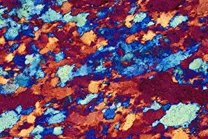
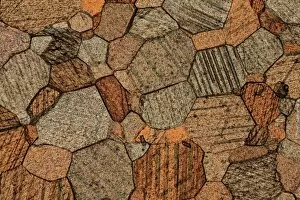
Polarised light micrograph of a thin section of quartzite, a metamorphic rock. The composition of quartzite varies: quartz is an essential component, with accessories of mica and feldspar

Background imageMagnified Image Collection: Marble, thin section, polarised LM

Marble, thin section, polarised LM
Polarised light micrograph of a sample of marble in thin section. Marble is a calcareous metamorphic rock, derived from fairly pure limestone

Background imageMagnified Image Collection: Salt crystals, light micrograph

Salt crystals, light micrograph
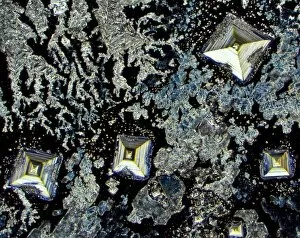
Salt. Dark field light micrograph of sodium chloride crystals, the common salt. This is a white crystalline compound widespread in nature as a solid (rock salt)

Background imageMagnified Image Collection: Insulin crystals, light micrograph

Insulin crystals, light micrograph
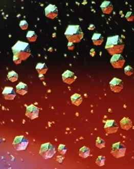
Insulin. Polarised light micrograph (PLM) of crystals of the hormone insulin. The crystals appear hexagonal in shape. The insulin molecule is made up of two chains of amino acids (A & B chains)

Background imageMagnified Image Collection: Purkinje nerve cells, light micrograph

Purkinje nerve cells, light micrograph
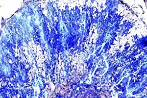
Purkinje nerve cells. Light micrograph of a section through a row of Purkinje nerve cells from the brains cerebellum. Each Purkinje cell is composed of a flask-shaped cell body which branch numerous

Background imageMagnified Image Collection: Silver Nitrate, light micrograph

Silver Nitrate, light micrograph
Composite crystals of Silver Nitrate, polarised light micrograph

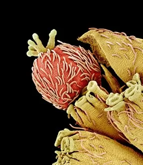
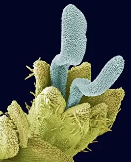
Background imageMagnified Image Collection: Euphorbia flower reproductive parts, SEM

Euphorbia flower reproductive parts, SEM
Euphorbia flower reproductive parts. Coloured scanning electron micrograph (SEM) of the reproductive parts of a Euphorbia supina flower

Background imageMagnified Image Collection: Polarised LM of vitamin D crystals

Polarised LM of vitamin D crystals
Vitamin D. Polarised light micrograph of crystals of vitamin D. Vitamin D is fat-soluble, and is found in foods such as animal fats, milk, butter and eggs

Background imageMagnified Image Collection: Polarised LM of niacinamide crystals

Polarised LM of niacinamide crystals
Nicotinamide. Polarised light micrograph of crystals of nicotinamide, or niacinamide. This is the amide of nicotinic acid. The acid and amide are both B vitamins, and are equally active

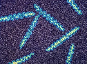
Background imageMagnified Image Collection: Treponema pallidum bacteria

Treponema pallidum bacteria

Background imageMagnified Image Collection: Gas gangrene bacteria, SEM

Gas gangrene bacteria, SEM
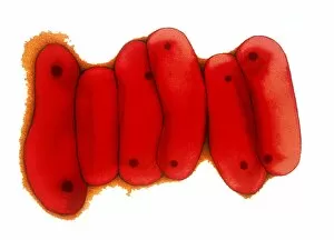
Gas gangrene bacteria (Clostridium perfringens), coloured scanning electron micrograph (SEM). These rod-shaped bacteria are pathogenic

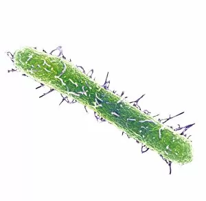
Background imageMagnified Image Collection: Salmonella typhimurium bacterium, SEM

Salmonella typhimurium bacterium, SEM
Salmonella typhimurium bacterium, coloured scanning electron micrograph (SEM). This bacterium is also known as Salmonella enterica serovar typhimurium

Background imageMagnified Image Collection: Spherical bacteria

Spherical bacteria. Computer artwork of chains of spherical bacteria (cocci). Typical cocci are Staphylococcus and Streptococcus bacteria, though there are many others as well

Background imageMagnified Image Collection: Tuberculosis bacteria, TEM

Tuberculosis bacteria, TEM
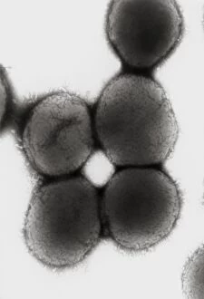
Tuberculosis bacteria. Coloured transmission electron micrograph (TEM) of a section through Mycobacterium tuberculosis bacteria

Background imageMagnified Image Collection: Moraxella catarrhalis bacteria

Moraxella catarrhalis bacteria

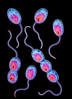
Background imageMagnified Image Collection: Rhodopseudomonas sp bacteria

Rhodopseudomonas sp bacteria

Background imageMagnified Image Collection: Anti-bacterial dressing

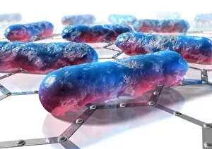
Anti-bacterial dressing. Computer artwork of rod- shaped bacteria (bacilli) on an anti-bacterial dressing. A typical design of such dressings is seen here

Background imageMagnified Image Collection: SEM of soil bacteria

SEM of soil bacteria

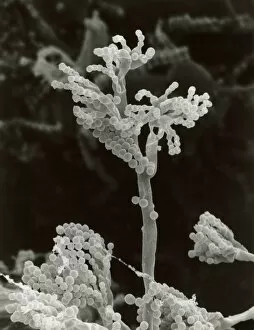
Background imageMagnified Image Collection: Penicillin fungus growing on cheddar cheese

Penicillin fungus growing on cheddar cheese
Scanning electron micrograph (SEM) the fruiting bodies or conidia of a penicillin fungus growing on a piece of English cheddar cheese which has gone mouldy in the refrigerator

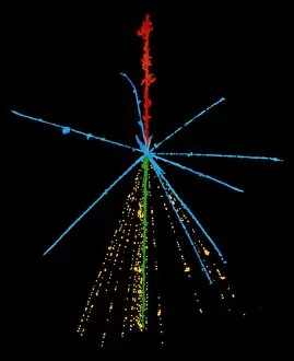
Background imageMagnified Image Collection: Emulsion photo of cosmic ray event

Emulsion photo of cosmic ray event
The power of a cosmic ray is shown in this false- colour emulsion photo of a cosmic ray sulphur nucleus (red) colliding with a nucleus in the emulsion

Background imageMagnified Image Collection: Particle tracks on a star chart

Particle tracks on a star chart

Background imageMagnified Image Collection: Pelargonium pollen grain, SEM

Pelargonium pollen grain, SEM
Pollen grain. Coloured scanning electron micrograph (SEM) of a grain of pollen from a geranium (Pelargonium sp.). Pollen grains contain the male reproductive cells of a plant

Background imageMagnified Image Collection: Disc floret, SEM

Disc floret, SEM
Disc floret. Coloured scanning electron micrograph (SEM) of a disc floret of a composite flower. Daisies (family Asteraceae, formerly Compositae) have inflorescences composed of two types of flowers

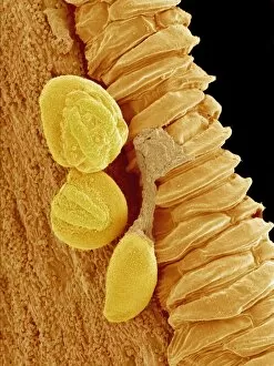
Background imageMagnified Image Collection: Gladiolus flower anther, SEM

Gladiolus flower anther, SEM
Gladiolus flower anther. Coloured scanning electron micrograph (SEM) of pollen grains (yellow) on a gladiolus flower (Gladiolus sp.) anther

Background imageMagnified Image Collection: Comfrey flower petal, SEM

Comfrey flower petal, SEM
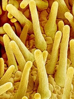
Comfrey flower petal. Coloured scanning electron micrograph (SEM) showing papillae on a petal from a comfrey (Symphytum officinale) flower. Papillae are projections from epidermal cells

Background imageMagnified Image Collection: Pollen grain surface, SEM

Pollen grain surface, SEM
Pollen grain surface. Coloured scanning electron micrograph (SEM) of the surface of a pollen grain. The characteristic outer wall, or exine

Background imageMagnified Image Collection: Spider lily flower stamen, SEM

Spider lily flower stamen, SEM
Spider lily flower stamen. Coloured scanning electron micrograph (SEM) of a cilia (hair-like structures) from a spider lily flower (Hymenocallis sp.) stamen

Background imageMagnified Image Collection: Open stoma, SEM

Open stoma, SEM
Open stoma. Coloured scanning electron micrograph (SEM) of an open stoma on the surface of a tobacco leaf (Nicotiana tabacum)

Background imageMagnified Image Collection: Light micrograph of vitamin B6 crystals

Light micrograph of vitamin B6 crystals
Polarised light micrograph of crystals of vitamin B6, known as Pyridoxine. Magnification: x63 at 35mm size

Background imageMagnified Image Collection: Clostridium tetani bacterium

Clostridium tetani bacterium
False-colour transmission electron micrograph (TEM) of the bacterium Clostridium tetani, a Gram- positive, spore-forming, anaerobic bacillus (rod-shaped bacterium) that causes tetanus

Background imageMagnified Image Collection: Germ, conceptual computer artwork

Germ, conceptual computer artwork


For sale as Licensed Images

Choose your image, Select your licence and Download the media

"Exploring the unseen wonders of the microscopic world: from particle tracks to intricate tissues and beyond. " "Unveiling the hidden beauty within: a closer look at cerebellum tissue through a mesmerizing light micrograph. " "A captivating glimpse into the mysteries of subatomic particles: behold the bubble chamber photo capturing sigma particle decay. " "The groundbreaking moment in science history: witness the first observation of omega-minus particle, forever changing our understanding of matter. " "Awe-inspiring beginnings: marvel at the intricate structure of a human blastocyst, where life takes its first steps. " "Diving deep into neural networks: an illuminating immunofluorescent LM reveals stunning neurons and astrocytes in all their glory. " "Unraveling nature's cycle within us: witness the uterus lining during menstruation, as seen through SEM imaging - a fascinating display of renewal and change. " "Peering into viral threats that loom above us all: explore avian flu virus up close, revealing its complex structure and potential dangers it holds. " "The crystalline beauty behind love and bonding: discover oxytocin hormone crystals under PLM C016 / 7196 microscopy - nature's secret ingredient for connection. " "A tapestry woven by nature itself: delve into intricately detailed fabric structures captured through SEM imaging - artistry on a microscopic scale. " "Glimpsing into windows to our souls with breathtaking precision: explore the iris of an eye like never before, revealing unique patterns that make each person truly one-of-a-kind. " "Revealing fragility beneath strength: examine osteoporotic bone under SEM, shedding light on this silent disease affecting millions worldwide.