Magnified Image Collection (page 9)

"Exploring the unseen wonders of the microscopic world: from particle tracks to intricate tissues and beyond

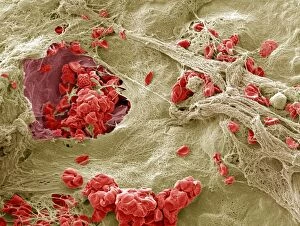
Background imageMagnified Image Collection: Healing wound, SEM

Healing wound, SEM
Healing skin wound, coloured scanning electron micrograph (SEM). A blood vessel is leaking at left. At right, red blood cells (or erythrocytes)

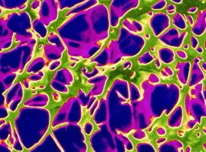
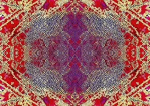
Background imageMagnified Image Collection: Computer artwork of osteoporosis affecting bone

Computer artwork of osteoporosis affecting bone
Osteoporosis. Computer artwork of a Scanning Electron Micrograph showing the effect of osteoporosis on bone tissue. This section through one of the vertebrae of the backbone shows the cancellous

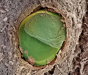
Background imageMagnified Image Collection: Embolism

Embolism. Coloured scanning electron micrograph (SEM) of a blockage, or embolism, in a blood vessel. Embolisms, particularly when they occur in vital organs such as the lungs (pulmonary embolism)

Background imageMagnified Image Collection: Coloured TEM of Tobacco Mosaic Viruses (TMV)

Coloured TEM of Tobacco Mosaic Viruses (TMV)
Tobacco mosaic viruses. Coloured transmission electron micrograph of a cluster of rod-shaped red virus particles of tobacco mosaic virus (TMV)

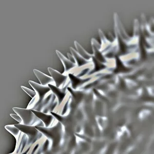
Background imageMagnified Image Collection: Influenza virus protein spikes

Influenza virus protein spikes. Computer artwork of part of the surface of a virus particle of the influenza (flu) virus. These spikes (formed by the surface proteins)

Background imageMagnified Image Collection: Influenza viruses

Influenza viruses. Coloured transmission electron micrograph (TEM) of influenza (flu) viruses. The flu virus belongs to the orthomyxovirus group of viruses which have an affinity for mucus

Background imageMagnified Image Collection: Coloured TEM of HHV6 infecting a cell

Coloured TEM of HHV6 infecting a cell
Coloured transmission electron micrograph of Human Herpes Virus type 6 (HHV6) infecting a human cell. Within a pseudopodium (cell outgrowth centre)

Background imageMagnified Image Collection: Angora wool

Angora wool fibres. Coloured scanning electron micrograph of fibres of angora wool. These show the overlapping scales typical of animal hairs

Background imageMagnified Image Collection: Cells

Cells. Computer artwork of non-specific cells. The red dot in the centre of each cell is the nucleus where the DNA (deoxyribonucleic acid) of each cell is stored

Background imageMagnified Image Collection: Graphite pencil core, SEM

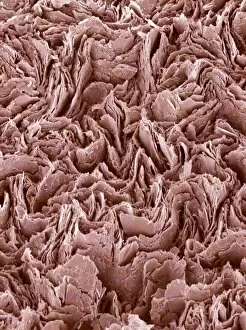
Graphite pencil core, SEM
Graphite pencil core. Coloured scanning electron micrograph (SEM) of layers making up the core of a graphite pencil. This core is made up of graphite

Background imageMagnified Image Collection: Human embryonic stem cell, SEM

Human embryonic stem cell, SEM
Stem cell. Coloured scanning electron micrograph (SEM) of a human embryonic stem cell (HESC, blue). HESCs are pluripotent

Background imageMagnified Image Collection: Embryonic stem cell, SEM

Embryonic stem cell, SEM
Embryonic stem cell. Coloured scanning electron micrograph (SEM) of a mouse embryonic stem cell. Embryonic stem cells are pluripotent

Background imageMagnified Image Collection: Stressed culture cell, SEM

Stressed culture cell, SEM

Background imageMagnified Image Collection: Watch gears, SEM

Watch gears, SEM
Watch gears, coloured scanning electron micrograph (SEM). These cogs move the hands of the watch

Background imageMagnified Image Collection: Pancreas cell, SEM

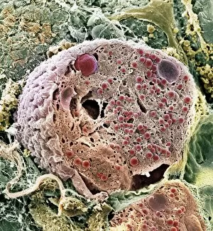
Pancreas cell, SEM
Pancreas cell. Coloured scanning electron micrograph (SEM) of an acinar (exocrine) pancreatic cell. Acinar cells produce and excrete digestive enzymes to the small intestine

Background imageMagnified Image Collection: SEM of snow & ice crystals

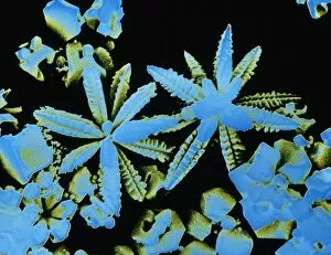
SEM of snow & ice crystals
False-colour scanning electron micrograph of a pair of tiny, flower-like ice crystals. Snow & ice crystals have a characteristic hexagonal symmetry

Background imageMagnified Image Collection: Climbing lily pollen, SEM

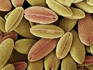
Climbing lily pollen, SEM
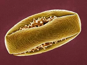
Climbing lily pollen grains. Coloured scanning electron micrograph (SEM) of pollen grains from the climbing lily flower (Gloriosa superba)

Background imageMagnified Image Collection: Horse chestnut pollen, SEM

Horse chestnut pollen, SEM
Horse chestnut pollen grain. Coloured scanning electron micrograph (SEM) of a pollen grain from a flowering horse chestnut tree (Aesculus hippocastanum)

Background imageMagnified Image Collection: Oestradiol, female sex hormone

Oestradiol, female sex hormone
Female sex hormone. Polarised light micrograph of crystals of Oestradiol, also known as beta- Estradiol. This is the most potent of the six naturally-occurring oestrogens

Background imageMagnified Image Collection: Serotonin crystals, light micrograph

Serotonin crystals, light micrograph
Serotonin. Polarised light micrograph of serotonin, a neurotransmitter. Serotonin is derived from tryptophan, and is found in the vertebrate brain

Background imageMagnified Image Collection: Balancing stone from inner ear, SEM

Balancing stone from inner ear, SEM
Balancing stone from inner ear. Coloured scanning electron micrograph (SEM) of crystals of calcium carbonate on the surface of a human otolith

Background imageMagnified Image Collection: Syenite, thin section, polarized LM

Syenite, thin section, polarized LM
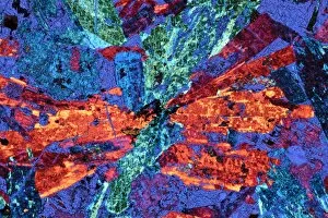
Polarised light micrograph of a thin section of syenite, a coarse-grained igneous rock, containing the mineral augite. Augite occurs as a prismatic crystal which in cross-section appears almost

Background imageMagnified Image Collection: Mica schist, thin section, polarised LM

Mica schist, thin section, polarised LM
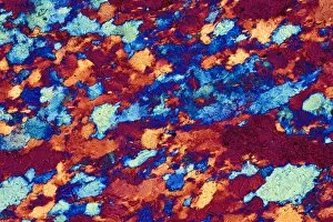
Polarised light micrograph of a thin section of mica schist, a type of metamorphic rock. Magnification: x12 at 35mm size

Background imageMagnified Image Collection: Limestone, thin section, polarised LM

Limestone, thin section, polarised LM
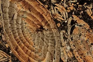
Nummulitic limestone. Polarised light micrograph of a thin section of a Nummulitic limestone. The large coloured objects are the remains of large foraminifera, Nummulites sp

Background imageMagnified Image Collection: Quartzite, thin section, polarized LM

Quartzite, thin section, polarized LM
Polarised light micrograph of a thin section of quartzite, a metamorphic rock. The composition of quartzite varies: quartz is an essential component, with accessories of mica and feldspar

Background imageMagnified Image Collection: Marble, thin section, polarised LM

Marble, thin section, polarised LM
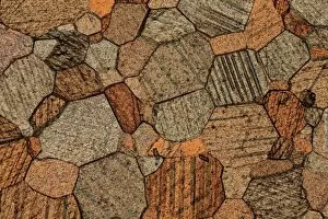
Polarised light micrograph of a sample of marble in thin section. Marble is a calcareous metamorphic rock, derived from fairly pure limestone

Background imageMagnified Image Collection: Salt crystals, light micrograph

Salt crystals, light micrograph
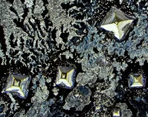
Salt. Dark field light micrograph of sodium chloride crystals, the common salt. This is a white crystalline compound widespread in nature as a solid (rock salt)

Background imageMagnified Image Collection: Insulin crystals, light micrograph

Insulin crystals, light micrograph
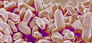
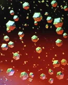
Insulin. Polarised light micrograph (PLM) of crystals of the hormone insulin. The crystals appear hexagonal in shape. The insulin molecule is made up of two chains of amino acids (A & B chains)

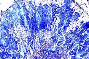
Background imageMagnified Image Collection: Purkinje nerve cells, light micrograph

Purkinje nerve cells, light micrograph
Purkinje nerve cells. Light micrograph of a section through a row of Purkinje nerve cells from the brains cerebellum. Each Purkinje cell is composed of a flask-shaped cell body which branch numerous

Background imageMagnified Image Collection: Silver Nitrate, light micrograph

Silver Nitrate, light micrograph
Composite crystals of Silver Nitrate, polarised light micrograph

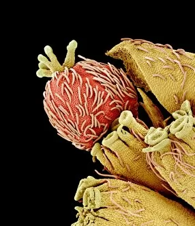
Background imageMagnified Image Collection: Euphorbia flower reproductive parts, SEM

Euphorbia flower reproductive parts, SEM
Euphorbia flower reproductive parts. Coloured scanning electron micrograph (SEM) of the reproductive parts of a Euphorbia supina flower

Background imageMagnified Image Collection: Polarised LM of vitamin D crystals

Polarised LM of vitamin D crystals
Vitamin D. Polarised light micrograph of crystals of vitamin D. Vitamin D is fat-soluble, and is found in foods such as animal fats, milk, butter and eggs

Background imageMagnified Image Collection: Polarised LM of niacinamide crystals

Polarised LM of niacinamide crystals
Nicotinamide. Polarised light micrograph of crystals of nicotinamide, or niacinamide. This is the amide of nicotinic acid. The acid and amide are both B vitamins, and are equally active

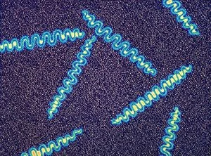
Background imageMagnified Image Collection: Treponema pallidum bacteria

Treponema pallidum bacteria


For sale as Licensed Images

Choose your image, Select your licence and Download the media

"Exploring the unseen wonders of the microscopic world: from particle tracks to intricate tissues and beyond. " "Unveiling the hidden beauty within: a closer look at cerebellum tissue through a mesmerizing light micrograph. " "A captivating glimpse into the mysteries of subatomic particles: behold the bubble chamber photo capturing sigma particle decay. " "The groundbreaking moment in science history: witness the first observation of omega-minus particle, forever changing our understanding of matter. " "Awe-inspiring beginnings: marvel at the intricate structure of a human blastocyst, where life takes its first steps. " "Diving deep into neural networks: an illuminating immunofluorescent LM reveals stunning neurons and astrocytes in all their glory. " "Unraveling nature's cycle within us: witness the uterus lining during menstruation, as seen through SEM imaging - a fascinating display of renewal and change. " "Peering into viral threats that loom above us all: explore avian flu virus up close, revealing its complex structure and potential dangers it holds. " "The crystalline beauty behind love and bonding: discover oxytocin hormone crystals under PLM C016 / 7196 microscopy - nature's secret ingredient for connection. " "A tapestry woven by nature itself: delve into intricately detailed fabric structures captured through SEM imaging - artistry on a microscopic scale. " "Glimpsing into windows to our souls with breathtaking precision: explore the iris of an eye like never before, revealing unique patterns that make each person truly one-of-a-kind. " "Revealing fragility beneath strength: examine osteoporotic bone under SEM, shedding light on this silent disease affecting millions worldwide.