
Maxilla Collection (#2)
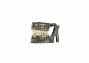
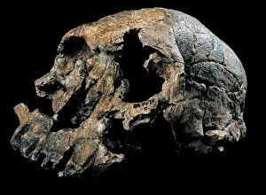
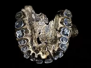
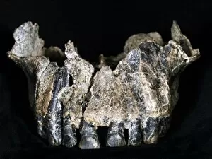
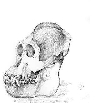
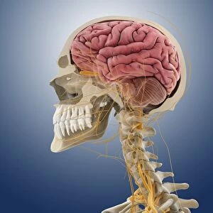
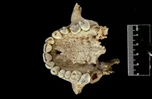
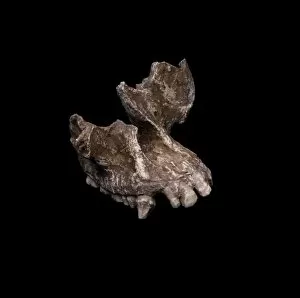
"Exploring the Intricacies of the Maxilla: A Journey into Skull Anatomy" Step into the world of Leonardo da Vinci's artwork as he meticulously studies skull anatomy


For sale as Licensed Images
Choose your image, Select your licence and Download the media
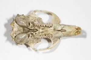
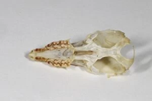
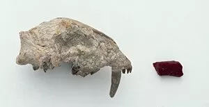
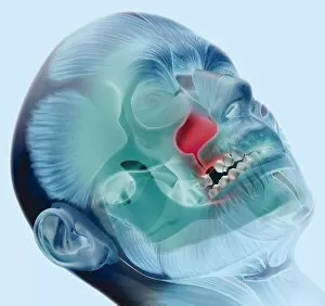
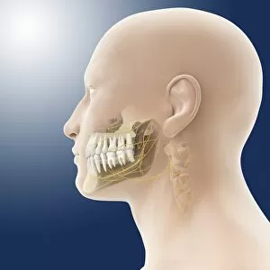
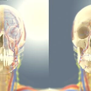
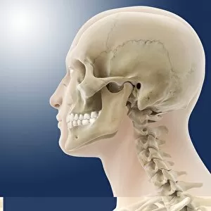
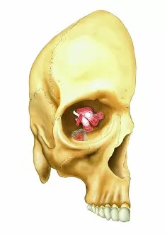
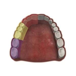
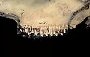
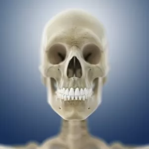
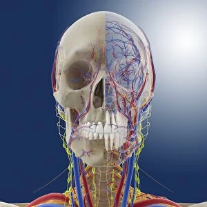
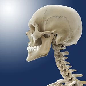
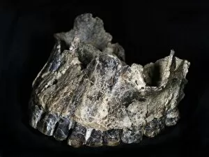
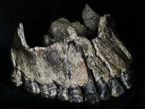
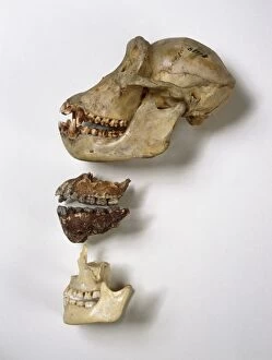
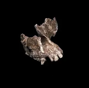
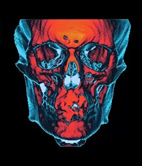
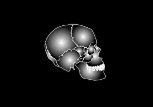
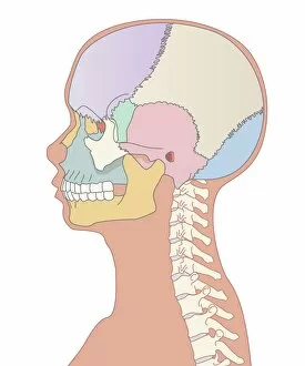
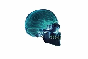
"Exploring the Intricacies of the Maxilla: A Journey into Skull Anatomy" Step into the world of Leonardo da Vinci's artwork as he meticulously studies skull anatomy, including the fascinating structure known as the maxilla. This captivating bone, located in both head and neck anatomy, holds a significant role in shaping our facial features. From different angles, we delve deep into understanding the complexities of human skull anatomy. With Leonardo's expertise guiding us, we examine every detail of the mouth cavity and its connection to this vital bone. As we explore further, even ancient species like Paranthropus boisei's cranium named Dear Boy captivate our attention. In a ghostly effect side view of a male human head with a visible skull, we witness how intricately intertwined these structures are. An anterior view provides labeled insights into each component while an inferior view showcases the base of the skull with informative labels. A unique perspective emerges when half muscles and half skull are displayed on a male human head—an artistic representation that highlights both strength and fragility. Additionally, a side view reveals an exploded version where individual parts come alive before our eyes. Through advanced technology like 3D rendering techniques, we gain new perspectives on how these bones interact within our bodies. The vertebral column harmoniously connects to form an awe-inspiring whole with the intricate architecture of our skulls at its pinnacle. As we immerse ourselves in this journey through time and artistry, it becomes clear that studying maxilla is not merely about bones but about unraveling mysteries hidden within humanity itself. So let us embrace this perspective—a window into our past—wherein lies knowledge waiting to be discovered anew.