
Medullary Ray Collection

"Unveiling the Intricate Medullary Ray

Background imageMedullary Ray Collection: African teak woody stem, light micrograph

African teak woody stem, light micrograph
African teak woody stem. Polarised light micrograph of a section through the stem of an African teak (Pericopsis elata) plant, showing its diffuse-porous xylem tissue

Background imageMedullary Ray Collection: Beech tree stem, light micrograph

Beech tree stem, light micrograph
Beech tree stem. Polarised light micrograph of a section through the woody stem of a common beech tree (Fagus sylvatica), showing diffuse-porous xylem vessels (dark blue)

Background imageMedullary Ray Collection: African mahogany stem, light micrograph

African mahogany stem, light micrograph
African mahogany stem. Polarised light micrograph of a section through the stem of an African mahogany (Khaya sp.) tree. The multicoloured cells are fibres, and fibre tracheids, which carry water

Background imageMedullary Ray Collection: African teak stem, light micrograph

African teak stem, light micrograph
African teak stem. Light micrograph of a section through the woody stem of an African teak (Pericopsis elata) plant, showing its diffuse-porous xylem tissue

Background imageMedullary Ray Collection: Cotton plant root, light micrograph

Cotton plant root, light micrograph
Cotton plant root. Light micrograph of a transverse section through a root of the cotton plant (Gossypium hirsutum). The layers from outer to centre (some very thin)

Background imageMedullary Ray Collection: Stinging nettle stem, light micrograph

Stinging nettle stem, light micrograph
Stinging nettle stem. Polarised light micrograph of a transverse section through a stem of the stinging nettle plant (Urtica dioica)

Background imageMedullary Ray Collection: Goosefoot root, light micrograph

Goosefoot root, light micrograph
Goosefoot root. Light micrograph of a section through the root of a goosefoot (Cheopodium sp.) plant. Xylem cells, which transport water around the plant are red

Background imageMedullary Ray Collection: Squash root, light micrograph

Squash root, light micrograph
Squash root. Light micrograph of a transverse section through a squash (Cucubita sp.) root. The large vessels (red) are xylem cells, which transport water. Parenchyma tissue is blue

Background imageMedullary Ray Collection: Elder tree stem, light micrograph

Elder tree stem, light micrograph
Elder tree stem. Light micrograph of a transverse section through the very young woody stem of an elder (Sambuca nigra) tree

Background imageMedullary Ray Collection: Mahogany wood structure, light micrograph

Mahogany wood structure, light micrograph
Mahogany wood structure. Polarised light micrograph of a longitudinal tangential section through a woody stem (xylem) of a mahogany (Swietenia sp.) tree


For sale as Licensed Images

Choose your image, Select your licence and Download the media

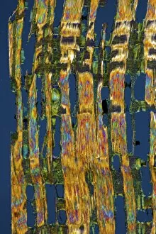
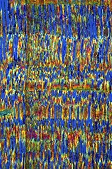
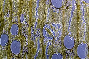
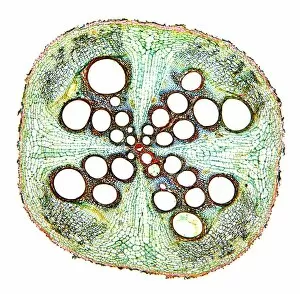
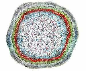
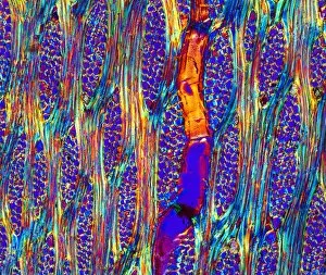
"Unveiling the Intricate Medullary Ray: A Journey into the Microscopic World of Tree Stems and Roots" Step into the fascinating realm of tree anatomy as we explore the medullary ray, a hidden wonder within beech, African teak, African mahogany, pine tree stems, and even cotton plant roots. Captured through light micrographs, these images offer a glimpse into the intricate structures that support these botanical giants. In our first encounter with beech tree stem's light micrograph, we witness delicate medullary rays branching out like ethereal sunbeams. These rays serve as highways for nutrients and water to travel vertically throughout the stem. Moving forward in our exploration, another beech tree stem unveils its secrets under microscopic scrutiny. The medullary ray takes center stage once again; its radial arrangement resembling an elegant tapestry woven by nature's hand. Venturing beyond beech trees' domain, we stumble upon an African teak woody stem's light micrograph. Here too lies evidence of medullary rays at work – their presence providing strength and stability to this majestic timber species. Continuing our journey through Africa's diverse flora kingdom, we encounter not one but two glimpses into African mahogany stems' inner world. In both instances captured by light micrographs, intricate patterns emerge as if painted onto canvas by a master artist - showcasing nature's unparalleled craftsmanship. Returning to African teak stems' embrace once more reveals yet another captivating sight under magnification. Medullary rays intertwine harmoniously with other cellular structures - a testament to their vital role in supporting life within this remarkable species. As our expedition nears its end, we turn our attention towards pine trees' sturdy stems. Light micrographs expose their unique medullary ray arrangements - reminiscent of interlocking fingers holding hands in unity against external forces.