Membrane Collection (#10)

The intricate anatomy of the human ear is beautifully depicted in this lithograph, published in 1876

Background imageMembrane Collection: Cell nucleus, TEM

Cell nucleus, TEM
Cell nucleus. Coloured transmission electron micrograph (TEM) of a section through a cell, showing the nucleus (large, spherical), and mitochondria (green)
Background imageMembrane Collection: Cryptochrome and rhodopsin pigments

Cryptochrome and rhodopsin pigments. Rhodopsin is a pigment found in the rod photoreceptor cells in the retina of the human eye, where it absorbs light falling on the retina

Background imageMembrane Collection: Outer membrane receptor protein molecule

Outer membrane receptor protein molecule
Outer membrane receptor protein. Molecular model of FecA an outer membrane receptor protein

Background imageMembrane Collection: Common pheasant male

Common pheasant male
Head of adult male common pheasant showing wattles and displaying the nictitating eye membrane. Dorset, UK March 2011

Background imageMembrane Collection: Cytoskeleton and membrane, diagram

Cytoskeleton and membrane, diagram
Cytoskeleton and membrane. Diagram showing the various structures associated with the cytoskeleton, the protein scaffolding found within cells

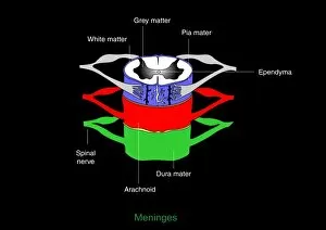
Background imageMembrane Collection: Meninges, artwork

Meninges, artwork
Meninges. Computer artwork showing the meninges surrounding the spinal cord. The meninges are the three membranes that envelop the brain and spinal cord

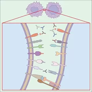
Background imageMembrane Collection: Immune system, artwork

Immune system, artwork
Immune system. Computer artwork showing how T-cells (right), antibodies (Y-shaped) and antigen-presenting cells (APC, left) interact during an immune response

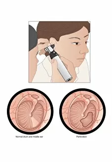
Background imageMembrane Collection: Perforated eardrum, artwork

Perforated eardrum, artwork
Perforated eardrum. Artwork of a perforated (torn) eardrum (right), or tympanic membrane, and a normal eardrum (left), as seen from the outer ear

Background imageMembrane Collection: Synapse structure, artwork

Synapse structure, artwork
Synapse structure. Cutaway artwork showing the structure of a synapse, the point where two nerve ending meet. The electrical impulse moving along a nerve is transmitted to the adjacent nerve by

Background imageMembrane Collection: Bacterial endospore formation, diagram

Bacterial endospore formation, diagram. The initial stage is at upper left, following the arrow to a ninth stage at lower right

Background imageMembrane Collection: Chloroplast structures, artwork

Chloroplast structures, artwork
Chloroplast structures. Cutaway artwork showing the internal structure of a chloroplast, the organelle in plant cells responsible for photosynthesis

Background imageMembrane Collection: Aquaporins, artwork

Aquaporins, artwork
Aquaporins. Computer artwork of water molecules (red and white) passing through pores (yellow) in a cell membrane (blue). These pores are proteins known as aquaporins that are embedded in

Background imageMembrane Collection: Aquaporin roles in the body

Aquaporin roles in the body. Aquaporins are membrane proteins that form channels (lower right) that help water molecules pass in and out of cells

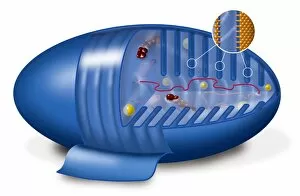
Background imageMembrane Collection: Mitochondrion, artwork

Mitochondrion, artwork
Mitochondrion. Cutaway artwork showing the internal structure of a mitochondrion. This structure, found in eukaryotic cells, is the site of energy production

Background imageMembrane Collection: Animal cell processes, artwork

Animal cell processes, artwork
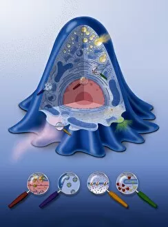
Animal cell processes. Cutaway artwork showing the structures inside an animal cell and four different processes that take place inside it or on its membrane (all marked by magnifying glasses)

Background imageMembrane Collection: Animal cell structure, artwork

Animal cell structure, artwork
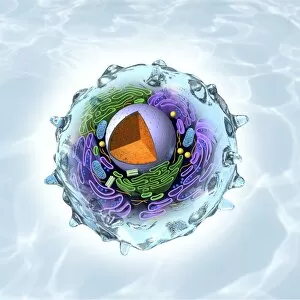
Animal cell structure. Artwork showing the internal structure of an animal cell. Inside the cell, the cellular structures (organelles) include the Golgi apparatus (green)

Background imageMembrane Collection: Multiverse layers, artwork

Multiverse layers, artwork. The multiverse, also called the meta-universe or metaverse, is the hypothetical set of possible universes, and includes all of space and time

Background imageMembrane Collection: Golgi apparatus, artwork

Golgi apparatus, artwork. This structure is an organelle found within eukaryotic cells. It receives proteins and lipids that are synthesised elsewhere on the endoplasmic reticulum

Background imageMembrane Collection: Desalination Reverse Osmosis filter

Desalination Reverse Osmosis filter
Desalination plant. A bank of Reverse Osmosis membrane filters. This facility turns salt water into drinking water using the Reverse Osmosis Process

Background imageMembrane Collection: Bacterial cell wall comparison, artwork

Bacterial cell wall comparison, artwork
Bacterial cell wall comparison. Computer artwork comparing the structure of the cell wall from a gram-negative bacterium (left) with that of a gram-positive bacterium (right)

Background imageMembrane Collection: Domestic greenhouse under construction

Domestic greenhouse under construction. The greenhouse structure is mounted on a base, complete with weed control fabric, but is unglazed

Background imageMembrane Collection: Domestic greenhouse in garden

Domestic greenhouse in garden
Domestic greenhouse under construction. The greenhouse is glazed and the base is lined with weed control fabric. There is no staging

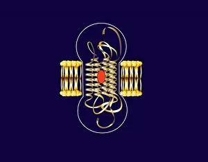
Background imageMembrane Collection: Bacteriorhodopsin, diagram

Bacteriorhodopsin, diagram
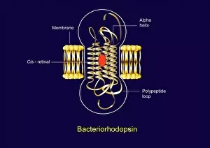
Bacteriorhodopsin. Diagram of the molecular structure of bacteriorhodopsin, a protein found in primitive micro-organisms known as Archaea

Background imageMembrane Collection: Bacteriorhodopsin, artwork

Bacteriorhodopsin, artwork
Bacteriorhodopsin. Artwork of the molecular structure of bacteriorhodopsin, a protein found in primitive micro-organisms known as Archaea

Background imageMembrane Collection: Muscle anatomy

Muscle anatomy. Cutaway artwork showing the anatomy and internal structure of a muscle, from the macroscopic (left) to the microscopic (right) level

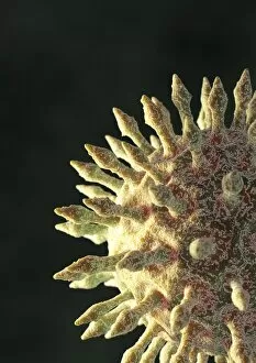
Background imageMembrane Collection: Virus particle, artwork

Virus particle, artwork. The spikes on this virus particle are surface proteins that help the particle attach to cell membranes

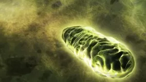
Background imageMembrane Collection: Mitochondrial energy, conceptual image

Mitochondrial energy, conceptual image. Computer artwork of a glowing mitochondrion, representing the energy produced by these cell organelles

Background imageMembrane Collection: Small intestine lining, light micrograph

Small intestine lining, light micrograph
Small intestine lining. Light micrograph of a section through the finger-like projections (villi) of the duodenum, the uppermost part of the small intestine

Background imageMembrane Collection: Mature poppy ovary, light micrograph

Mature poppy ovary, light micrograph
Mature poppy ovary. Light micrograph (LM) of a cross-section through a mature poppy ovary, which is incompletely divided by septa (membranes) that do not meet in the centre

Background imageMembrane Collection: Influenza virus structure, artwork

Influenza virus structure, artwork
Influenza virus structure, cutaway artwork. The core of the virus is its genetic material, here 8 coloured ribbons of single-stranded RNA (ribonucleic acid)

Background imageMembrane Collection: Orchid tuber, light micrograph

Orchid tuber, light micrograph
Orchid tuber. Polarised light micrograph of a transverse section through the root tuber of an orchid (Phaius grandifolius) showing starch grains (blue-red and yellow-green)

Background imageMembrane Collection: ZP3 glycoprotein molecule

ZP3 glycoprotein molecule
ZP3 glycoprotein. Computer artwork of a molecule of ZP3 (zona pellucida glycoprotein 3), a sperm receptor in the external membrane (zona pellucida) of a female germ cell (egg)

Background imageMembrane Collection: ATPase muscle enzyme

ATPase muscle enzyme
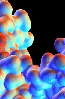
Calcium pumping ATPase enzyme. Computer model of an electrostatic potential surface map of part of the ATPase enzyme that pumps calcium in and out of muscle cells and controls muscle contractions

Background imageMembrane Collection: Horseshoe clam, Hippopus hippopus, Rongelap, Marshall Islands, Micronesia

Horseshoe clam, Hippopus hippopus, Rongelap, Marshall Islands, Micronesia
Andre Seale / SplashdownDirect

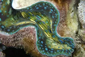
Background imageMembrane Collection: Fluted giant clam, Tridacna squamosa, Rongelap, Marshall Islands, Micronesia

Fluted giant clam, Tridacna squamosa, Rongelap, Marshall Islands, Micronesia
Andre Seale / SplashdownDirect

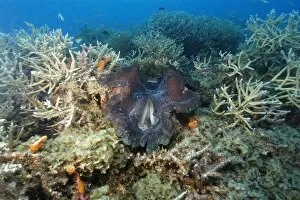
Background imageMembrane Collection: Giant clam, Tridacna gigas, Rongelap, Marshall Islands, Micronesia

Giant clam, Tridacna gigas, Rongelap, Marshall Islands, Micronesia
Andre Seale / SplashdownDirect

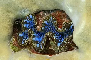
Background imageMembrane Collection: Small giant clam, Tridacna maxima, surrounded by lobe coral, Porites lutea, Namu atoll

Small giant clam, Tridacna maxima, surrounded by lobe coral, Porites lutea, Namu atoll, Marshall Islands (N. Pacific)
Andre Seale / SplashdownDirect

Background imageMembrane Collection: Fluted giant clam, Tridacna squamosa, Rongelap, Marshall Islands, Micronesia

Fluted giant clam, Tridacna squamosa, Rongelap, Marshall Islands, Micronesia
Andre Seale / SplashdownDirect

Background imageMembrane Collection: Horseshoe clam, Hippopus hippopus, Rongelap, Marshall Islands, Micronesia

Horseshoe clam, Hippopus hippopus, Rongelap, Marshall Islands, Micronesia
Andre Seale / SplashdownDirect

Background imageMembrane Collection: Bacterial cell

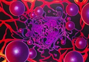
Bacterial cell, computer artwork. This is ageneralised bacterium, illustrating features ofdifferent bacterial types that would not becombined in a single cell


For sale as Licensed Images

Choose your image, Select your licence and Download the media

The intricate anatomy of the human ear is beautifully depicted in this lithograph, published in 1876. One notable feature highlighted here is the membrane, which plays a crucial role in transmitting sound waves to our auditory system. Moving on to cellular structures, the rough endoplasmic reticulum (ER) takes center stage under a transmission electron microscope (TEM). This network of membranes within cells is responsible for protein synthesis and transport. Artwork showcasing different cell types also emphasizes the significance of membranes. From the delicate cell membrane itself, represented by artwork C013 / 7467, to mitochondria seen through TEM imaging - these organelles possess their own unique membranes that regulate various cellular functions. Intriguingly, even chloroplasts have their own distinct membrane structure as revealed by artwork dedicated to studying photosynthesis. These specialized organelles found in plants are responsible for converting sunlight into energy. Beyond biology, membranes find relevance elsewhere too. Think about damp-proofing measures taken in houses – membranes act as barriers against moisture infiltration and protect our living spaces from potential damage. However, not all mentions of they are positive. Bacterial meningitis can be detected through MRI scans where inflammation affects the protective brain meninges' integrity. Understanding how pathogens breach these defensive layers helps diagnose and treat such infections effectively. Nature's wonders also exhibit fascinating adaptations involving membranes; take Plecotus sp. , commonly known as long-eared bats with their remarkable hearing abilities thanks to specialized ear membrane structures aiding echolocation skills. Zooming into finer details under TEM again reveals eye muscles' intricate arrangement (TEM C014 / 1468), highlighting how well-organized muscle fibers rely on precise membranous connections for coordinated movement and vision control. Lastly, let's not forget intestinal microvilli observed through TEM – finger-like projections covered by plasma membrane lining our intestines play a vital role in nutrient absorption during digestion processes. From ancient lithographs to modern imaging techniques, the significance of membranes spans across various fields.