Mitochondrion Collection (#4)

The mitochondrion, often referred to as the powerhouse of the cell, is a fascinating and vital organelle found in various cell types

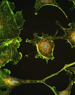
Background imageMitochondrion Collection: Human epithelial cells

Human epithelial cells. Fluorescent light micrograph of human epithelial cells in culture. The nuclei, which contain the cells genetic information DNA (deoxyribonucleic acid), have been dyed blue

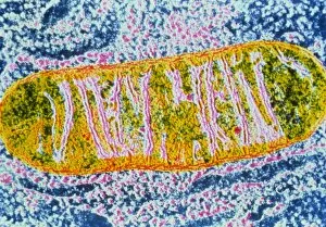
Background imageMitochondrion Collection: Cell mitochondrion

Cell mitochondrion
False-colour transmission electron micrograph of a mitochondrion from an unidentified cell. Mitochondria are the sites of cell respiration; the chemical process which uses molecular oxygen to oxidise

Background imageMitochondrion Collection: Animal cell structure, computer artwork

Animal cell structure, computer artwork
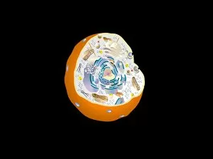
Animal cell. Computer artwork of a section through an animal cell. At the centre is the nucleus, which contains the cells genetic information in the form of DNA (deoxyribonucleic acid)

Background imageMitochondrion Collection: Cell organelles, TEM

Cell organelles, TEM
Cell organelles. Coloured transmission electron micrograph (TEM) of several organelles in an animal cell. At centre, the nucleus (dark purple, round) contains the cells genetic material (DNA)

Background imageMitochondrion Collection: Mitochondria

Mitochondria. Coloured transmission electron micrograph of mitochondria (circles). A mitochondrion is a membrane-bound organelle that exists in eukaryotic cells

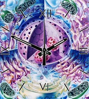
Background imageMitochondrion Collection: Cellular clock

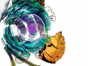
Cellular clock. Conceptual artwork of a clock and the internal structures (organelles) of an animal cell. This could represent the rate of metabolic cell reactions

Background imageMitochondrion Collection: Cardiac muscle, SEM

Cardiac muscle, SEM
Cardiac muscle. Coloured scanning electron micrograph (SEM) of a bundle of cardiac muscle fibrils (green) from a healthy heart. Mitochondria (round, orange) supply the muscle cells with energy

Background imageMitochondrion Collection: Mitochondrial Eve

Mitochondrial Eve. Conceptual computer artwork of a circular molecule of mitochondrial deoxyribonucleic acid (DNA) floating above a hand. Mitochondria are cell organelles that produce energy

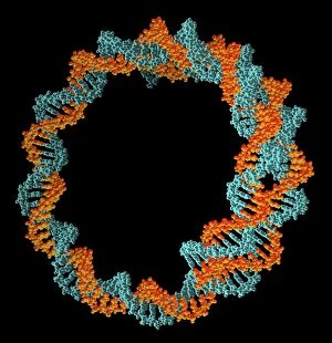
Background imageMitochondrion Collection: Circular DNA molecule, artwork

Circular DNA molecule, artwork
Circular DNA (deoxyribonucleic acid) molecule, computer artwork. Circular DNA has no ends, but consists of a ring structure

Background imageMitochondrion Collection: Cell nucleus, TEM

Cell nucleus, TEM
Cell nucleus. Coloured transmission electron micrograph (TEM) of a section through a cell, showing the nucleus (large, spherical), and mitochondria (green)

Background imageMitochondrion Collection: Muscle cell anatomy, artwork

Muscle cell anatomy, artwork
Muscle cell anatomy. Artwork of the anatomy of a muscle cell, also known as a muscle fibre. The cell is surrounded by a plasma membrane called the sarcolemma (grey)

Background imageMitochondrion Collection: Sperm cell, artwork

Sperm cell, artwork
Sperm cell. Artwork showing the structure of a sperm cell, or spermatozoon, the male sex cell. It consists of a tapered head (red) that contains the genetic material

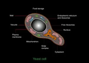
Background imageMitochondrion Collection: Yeast cell, artwork

Yeast cell, artwork
Yeast cell. Computer artwork showing the structure of a yeast cell

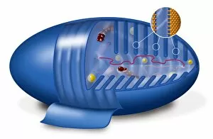
Background imageMitochondrion Collection: Mitochondrion, artwork

Mitochondrion, artwork
Mitochondrion. Cutaway artwork showing the internal structure of a mitochondrion. This structure, found in eukaryotic cells, is the site of energy production

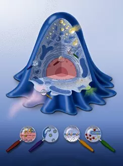
Background imageMitochondrion Collection: Animal cell processes, artwork

Animal cell processes, artwork
Animal cell processes. Cutaway artwork showing the structures inside an animal cell and four different processes that take place inside it or on its membrane (all marked by magnifying glasses)

Background imageMitochondrion Collection: Lung cells, fluorescent micrograph

Lung cells, fluorescent micrograph
Lung cells. Immunofluorescence light micrograph of pulmonary endothelial cells. Endothelial cells are specialized epithelial cells that line the inner surface of blood vessels

Background imageMitochondrion Collection: Retinal rod cell anatomy, diagram

Retinal rod cell anatomy, diagram

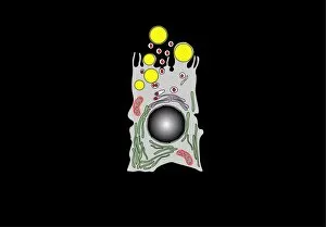
Background imageMitochondrion Collection: Milk-producing cell, artwork

Milk-producing cell, artwork
Milk-producing cell. Artwork of the anatomical structure of a lacteal (milk-producing) cell. Normal cell organelles include the nucleus (black, centre), the golgi apparatus (white)

Background imageMitochondrion Collection: Retinal rod cell anatomy, artwork

Retinal rod cell anatomy, artwork
Retinal rod cell anatomy. Artwork of the external (left) and internal (right) anatomical structure of a retinal cell. These cells are found in the retina that lines the back of the eyes

Background imageMitochondrion Collection: Milk-producing cell, diagram

Milk-producing cell, diagram
Milk-producing cell. Diagram of the anatomical structure of a lacteal (milk-producing) cell. Normal cell organelles include the nucleus (black, centre), the golgi apparatus (white)

Background imageMitochondrion Collection: Animal cell anatomy, artwork

Animal cell anatomy, artwork
Animal cell anatomy. Artwork showing the internal and external anatomy of an animal cell

Background imageMitochondrion Collection: Animal cell, artwork

Animal cell, artwork
Animal cell. Computer artwork of an expanded animal cell. At centre left is the nucleus (purple sphere), which contains the cells genetic information in the form of DNA (deoxyribonucleic acid)

Background imageMitochondrion Collection: Macrophage cells, TEM

Macrophage cells, TEM
Macrophage cells, coloured transmission electron micrograph (TEM). The cell nuclei are purple. Mitochondria (dark pink ovals) in the cytoplasm produce energy for the cell

Background imageMitochondrion Collection: Mitochondrial energy, conceptual image

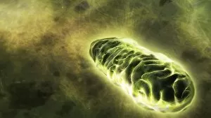
Mitochondrial energy, conceptual image. Computer artwork of a glowing mitochondrion, representing the energy produced by these cell organelles

Background imageMitochondrion Collection: Mitochondrion, SEM

Mitochondrion, SEM
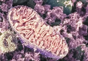
Mitochondrion. Coloured scanning electron micrograph (SEM) of a mitochondrion in a nerve cell. Mitochondria are a type of organelle found in the cytoplasm of eukaryotic cells

Background imageMitochondrion Collection: Cell organelles, SEM

Cell organelles, SEM
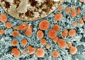
Cell organelles. Coloured scanning electron micrograph (SEM) of a section through a cell from a kidney proximal tubule. The cell nucleus (partially seen at top left)

Background imageMitochondrion Collection: Cell nucleus, SEM

Cell nucleus, SEM
Cell nucleus. Coloured scanning electron micrograph (SEM) of a section through the nucleus (centre right) of a kidney cell

Background imageMitochondrion Collection: RNA processing protein, molecular model

RNA processing protein, molecular model
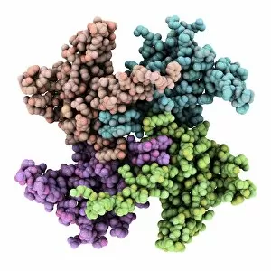
RNA processing protein, RNase MRP. Computer model showing the molecular structure of mitochondrial RNase MRP (mitochondrial RNA processing)

Background imageMitochondrion Collection: Intestinal smooth muscle cells, TEM

Intestinal smooth muscle cells, TEM
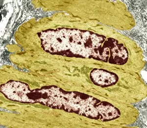
Intestinal smooth muscle cells. Coloured transmission electron micrograph (TEM) of a section through several smooth muscle cells from the intestines


For sale as Licensed Images

Choose your image, Select your licence and Download the media

The mitochondrion, often referred to as the powerhouse of the cell, is a fascinating and vital organelle found in various cell types. In budding yeast cells, it plays a crucial role in energy production through cellular respiration. This tiny structure can be visualized using transmission electron microscopy (TEM), revealing its intricate details. Not limited to yeast cells, mitochondria are present in different cell types throughout our bodies. TEM images showcase their distinct shapes and sizes within plasma cells, eye muscles, macrophage cells, and hepatocyte liver cells. These microscopic snapshots allow scientists to study their functions and understand how they contribute to overall cellular health. Scanning electron microscopy (SEM) provides another perspective on mitochondria's appearance within animal cell structures. These high-resolution images capture the surface of these organelles with stunning detail. Illustrations further enhance our understanding of mitochondria's complex composition. They depict an animal cell nucleus containing cytoplasm filled with numerous mitochondria units intertwined with DNA strands and chromosomes. Such illustrations also highlight TFAM transcription factors bound to DNA molecules—an essential process for regulating mitochondrial gene expression. Artwork showcasing muscle fiber structure emphasizes the significance of mitochondria in muscle function. Their abundance within muscle fibers ensures efficient energy supply during physical activity.