Mitochondrion Collection (#2)

The mitochondrion, often referred to as the powerhouse of the cell, is a fascinating and vital organelle found in various cell types

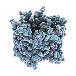
Background imageMitochondrion Collection: VDAC-1 ion channel protein C015 / 8249

VDAC-1 ion channel protein C015 / 8249
VDAC-1 ion channel protein, molecular model. This is the human voltage-dependent anion-selective channel protein 1 (VDAC-1)

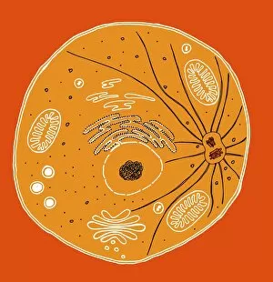
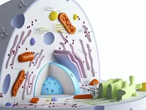
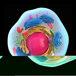
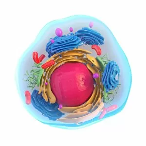
Background imageMitochondrion Collection: Animal cell, illustration C018 / 0734

Animal cell, illustration C018 / 0734
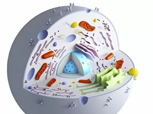
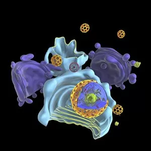
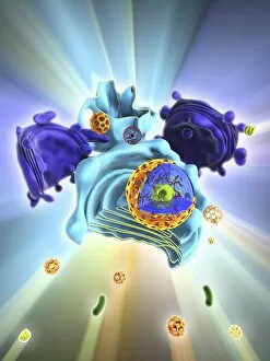
Animal cell. Illustration showing the organelles present in an animal cell. At lower centre is the nucleus, which contains the cells genetic information in the form of DNA (deoxyribonucleic acid)

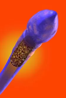
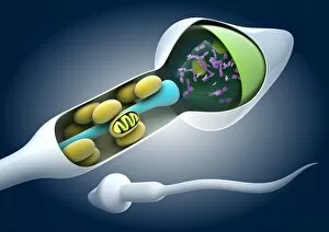
Background imageMitochondrion Collection: Sperm cell, artwork C018 / 6996

Sperm cell, artwork C018 / 6996
Sperm cell anatomy. Cutaway computer artwork showing the internal structure of a sperm cell (spermatozoon), the male sex cell
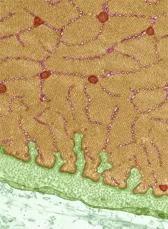
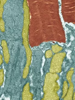
Background imageMitochondrion Collection: Eye muscle, TEM C014 / 1467

Eye muscle, TEM C014 / 1467
Eye muscle. Transmission electron micrograph (TEM) of a section through a striated muscle cell from the ciliary muscle of a human eye
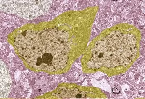
Background imageMitochondrion Collection: Intestinal gland cell, TEM C014 / 1442

Intestinal gland cell, TEM C014 / 1442
Intestinal gland cell. Transmission electron micrograph (TEM) of a section through an undifferentiated cell from the intestinal tract, showing extensive rough endoplasmic reticulum (ER)

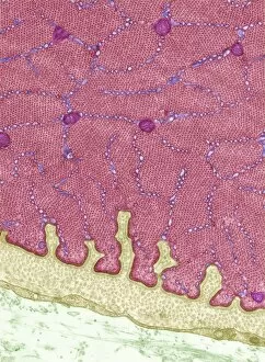
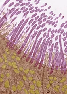
Background imageMitochondrion Collection: Trachea lining, TEM C014 / 1469

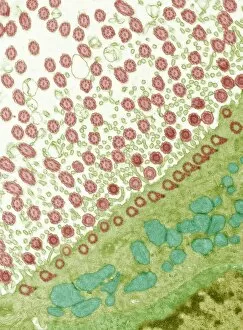
Trachea lining, TEM C014 / 1469
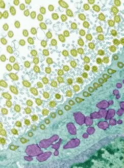
Trachea lining. Transmission electron micrograph (TEM) of a longitudinal section through the lining of the trachea (windpipe), which links the larynx (voicebox) to the lungs

Background imageMitochondrion Collection: Eye muscle, TEM C014 / 1466

Eye muscle, TEM C014 / 1466
Eye muscle. Transmission electron micrograph (TEM) of a section through a striated muscle cell from the ciliary muscle of a human eye

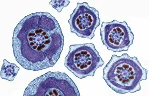
Background imageMitochondrion Collection: Sperm tails, TEM C014 / 1463

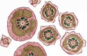
Sperm tails, TEM C014 / 1463
Sperm tails. Transmission electron micrograph (TEM) of sections through the tails of sperm (spermatozoa) cells at various levels

Background imageMitochondrion Collection: Trachea lining, TEM C014 / 1471

Trachea lining, TEM C014 / 1471
Trachea lining. Transmission electron micrograph (TEM) of a transverse section through the lining of the trachea (windpipe), which links the larynx (voicebox) to the lungs

Background imageMitochondrion Collection: Sperm tails, TEM C014 / 1465

Sperm tails, TEM C014 / 1465
Sperm tails. Transmission electron micrograph (TEM) of sections through the tails of sperm (spermatozoa) cells at various levels

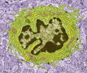
Background imageMitochondrion Collection: Monocyte white blood cell, TEM

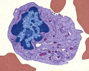
Monocyte white blood cell, TEM
Monocyte white blood cell. Transmission electron micrograph (TEM) of a section through a monocyte white blood cell, showing the cell nucleus (blue)

Background imageMitochondrion Collection: Sperm tails, TEM C014 / 1464

Sperm tails, TEM C014 / 1464
Sperm tails. Transmission electron micrograph (TEM) of sections through the tails of sperm (spermatozoa) cells at various levels

Background imageMitochondrion Collection: Trachea lining, TEM C014 / 1470

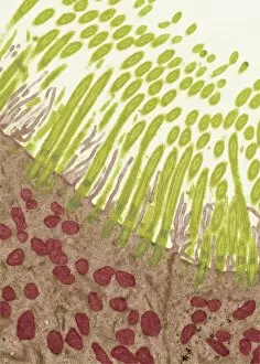
Trachea lining, TEM C014 / 1470
Trachea lining. Transmission electron micrograph (TEM) of a longitudinal section through the lining of the trachea (windpipe), which links the larynx (voicebox) to the lungs

Background imageMitochondrion Collection: Trachea lining, TEM C014 / 1472

Trachea lining, TEM C014 / 1472
Trachea lining. Transmission electron micrograph (TEM) of a transverse section through the lining of the trachea (windpipe), which links the larynx (voicebox) to the lungs

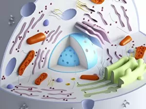
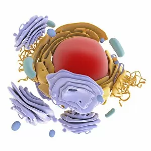
Background imageMitochondrion Collection: Animal cell organelles, artwork

Animal cell organelles, artwork
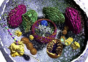
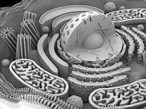
Animal cell organelles. Artwork showing the organelles in a eukaryotic cell. This is an animal cell. Structures include the nucleus (centre) which has a membrane with nuclear pores (purple)

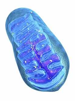
Background imageMitochondrion Collection: Mitochondrion structure, artwork

Mitochondrion structure, artwork
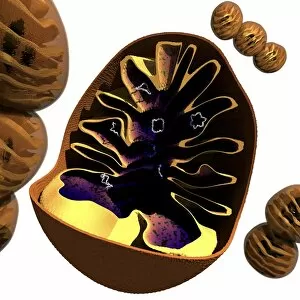
Mitochondrion structure. Artwork showing the internal structure of a mitochondrion. This structure, found in eukaryotic cells, is the site of energy production

Background imageMitochondrion Collection: Mitochondrial structure, artwork

Mitochondrial structure, artwork
Mitochondrial structure. Computer artwork of a section through a mitochondrion, showing the internal structure and a loop of mitochondrial DNA (deoxyribonucleic acid, mtDNA)

Background imageMitochondrion Collection: TFAM transcription factor bound to DNA C015 / 7060

TFAM transcription factor bound to DNA C015 / 7060
TFAM transcription factor bound to DNA, molecular model. Human mitochondrial transcription factor A (TFAM, pink) bound to a strand of DNA (deoxyribonucleic acid, orange and green)

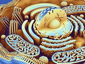
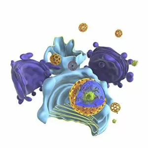
Background imageMitochondrion Collection: Animal cell organelles, artwork C015 / 6793

Animal cell organelles, artwork C015 / 6793
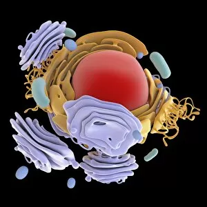
Animal cell organelles. Computer artwork showing the organelles in a eukaryotic cell. This is an animal cell. Structures include the nucleus (centre) which has a membrane with nuclear pores

Background imageMitochondrion Collection: Animal cell organelles, artwork C015 / 6792

Animal cell organelles, artwork C015 / 6792
Animal cell organelles. Computer artwork showing the organelles in a eukaryotic cell. This is an animal cell. Structures include the nucleus (centre) which has a membrane with nuclear pores

Background imageMitochondrion Collection: Mitochondrial structure, artwork C015 / 6784

Mitochondrial structure, artwork C015 / 6784
Mitochondrial structure. Computer artwork of a mitochondrion, showing a loop of mitochondrial DNA (deoxyribonucleic acid, mtDNA), the organelles genetic material

Background imageMitochondrion Collection: Animal cell organelles, artwork C015 / 6775

Animal cell organelles, artwork C015 / 6775
Animal cell organelles. Computer artwork showing the organelles in a eukaryotic cell. This is an animal cell. Structures include the nucleus (centre) which has a membrane with nuclear pores

Background imageMitochondrion Collection: Animal cell organelles, artwork C015 / 6778

Animal cell organelles, artwork C015 / 6778
Animal cell organelles. Computer artwork showing the organelles in a eukaryotic cell. This is an animal cell. Structures include the nucleus (centre) which has a membrane with nuclear pores

Background imageMitochondrion Collection: Animal cell organelles, artwork C015 / 6777

Animal cell organelles, artwork C015 / 6777
Animal cell organelles. Computer artwork showing the organelles in a eukaryotic cell. This is an animal cell. Structures include the nucleus (centre) which has a membrane with nuclear pores

Background imageMitochondrion Collection: Mitochondrial structure, artwork C015 / 6765

Mitochondrial structure, artwork C015 / 6765
Mitochondrial structure. Computer artwork of a mitochondrion, showing a loop of mitochondrial DNA (deoxyribonucleic acid, mtDNA), the organelles genetic material

Background imageMitochondrion Collection: Animal cell organelles, artwork C016 / 0619

Animal cell organelles, artwork C016 / 0619
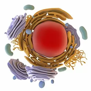
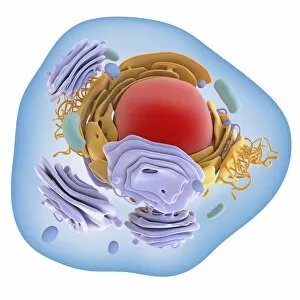
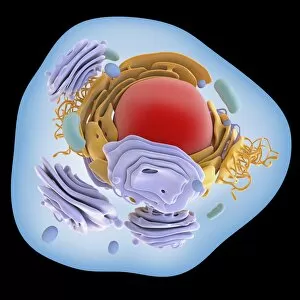
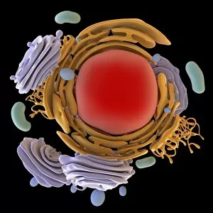
Computer artwork showing the organelles in a eukaryotic cell. This is an animal cell. Structures include the nucleus (centre, red) which has a membrane with nuclear pores

Background imageMitochondrion Collection: Animal cell organelles, artwork C016 / 0621

Animal cell organelles, artwork C016 / 0621
Computer artwork showing the organelles in a eukaryotic cell. This is an animal cell. Structures include the nucleus (centre, red) which has a membrane with nuclear pores

Background imageMitochondrion Collection: Animal cell organelles, artwork C016 / 0620

Animal cell organelles, artwork C016 / 0620
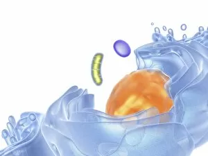
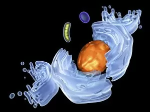
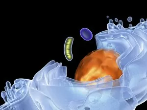
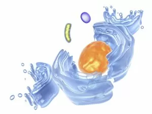
Computer artwork showing the organelles in a eukaryotic cell. This is an animal cell. Structures include the nucleus (centre, orange) which has a membrane with nuclear pores

Background imageMitochondrion Collection: Animal cell organelles, artwork C016 / 0617

Animal cell organelles, artwork C016 / 0617
Computer artwork showing the organelles in a eukaryotic cell. This is an animal cell. Structures include the nucleus (centre, red) which has a membrane with nuclear pores

Background imageMitochondrion Collection: Animal cell organelles, artwork C016 / 0618

Animal cell organelles, artwork C016 / 0618
Computer artwork showing the organelles in a eukaryotic cell. This is an animal cell. Structures include the nucleus (centre, orange) which has a membrane with nuclear pores

Background imageMitochondrion Collection: Animal cell organelles, artwork C016 / 0615

Animal cell organelles, artwork C016 / 0615
Computer artwork showing the organelles in a eukaryotic cell. This is an animal cell. Structures include the nucleus (centre, orange) which has a membrane with nuclear pores

Background imageMitochondrion Collection: Animal cell organelles, artwork C016 / 0616

Animal cell organelles, artwork C016 / 0616
Computer artwork showing the organelles in a eukaryotic cell. This is an animal cell. Structures include the nucleus (centre, orange) which has a membrane with nuclear pores

Background imageMitochondrion Collection: Animal cell organelles, artwork C016 / 0611

Animal cell organelles, artwork C016 / 0611
Computer artwork showing the organelles in a eukaryotic cell. This is an animal cell. Structures include the nucleus (centre, red) which has a membrane with nuclear pores

Background imageMitochondrion Collection: Animal cell organelles, artwork C016 / 0612

Animal cell organelles, artwork C016 / 0612
Computer artwork showing the organelles in a eukaryotic cell. This is an animal cell. Structures include the nucleus (centre, orange) which has a membrane with nuclear pores

Background imageMitochondrion Collection: Animal cell organelles, artwork C016 / 0605

Animal cell organelles, artwork C016 / 0605
Computer artwork showing the organelles in a eukaryotic cell. This is an animal cell. Structures include the nucleus (centre, red) which has a membrane with nuclear pores

Background imageMitochondrion Collection: Animal cell organelles, artwork C016 / 0610

Animal cell organelles, artwork C016 / 0610
Computer artwork showing the organelles in a eukaryotic cell. This is an animal cell. Structures include the nucleus (centre, orange) which has a membrane with nuclear pores

Background imageMitochondrion Collection: Animal cell organelles, artwork C016 / 0604

Animal cell organelles, artwork C016 / 0604
Computer artwork showing the organelles in a eukaryotic cell. This is an animal cell. Structures include the nucleus (centre, red) which has a membrane with nuclear pores

Background imageMitochondrion Collection: Animal cell organelles, artwork C016 / 0606

Animal cell organelles, artwork C016 / 0606
Computer artwork showing the organelles in a eukaryotic cell. This is an animal cell. Structures include the nucleus (centre, orange) which has a membrane with nuclear pores

Background imageMitochondrion Collection: Mitochondrial inclusions, TEM C016 / 5807

Mitochondrial inclusions, TEM C016 / 5807
Mitochondrial inclusions. Transmission electron micrograph (TEM) of a section through a mitochondrion showing crystalline inclusions (thick parallel rods) in the intermembrane spaces

Background imageMitochondrion Collection: Mitochondrial inclusions, TEM C016 / 5806

Mitochondrial inclusions, TEM C016 / 5806
Mitochondrial inclusions. Transmission electron micrograph (TEM) of a section through a mitochondrion showing crystalline inclusions (thick parallel rods) in the intermembrane spaces

Background imageMitochondrion Collection: Animal cell, artwork C013 / 9985

Animal cell, artwork C013 / 9985
Animal cell. Computer artwork showing the cell organelles found inside a typical animal cell. The nucleus (large round) can be seen at centre

Background imageMitochondrion Collection: Animal cell, artwork C013 / 9984

Animal cell, artwork C013 / 9984
Animal cell. Computer artwork showing the cell organelles found inside a typical animal cell. The nucleus (large round) can be seen at centre

Background imageMitochondrion Collection: Sperm cell anatomy, artwork C013 / 4648

Sperm cell anatomy, artwork C013 / 4648
Sperm cell anatomy. Cutaway computer artwork showing the internal structure of a sperm cell (spermatozoon), the male sex cell

Background imageMitochondrion Collection: Mitochondria, artwork C013 / 4993

Mitochondria, artwork C013 / 4993
Mitochondria, computer artwork. Mitochondria are a type of organelle found in the cytoplasm of eukaryotic cells. They oxidise sugars and fats to produce energy in a process called respiration

Background imageMitochondrion Collection: Mitochondria, artwork C013 / 4991

Mitochondria, artwork C013 / 4991
Mitochondria, computer artwork. Mitochondria are a type of organelle found in the cytoplasm of eukaryotic cells. They oxidise sugars and fats to produce energy in a process called respiration

Background imageMitochondrion Collection: Retinal rod cell, TEM C013 / 4805

Retinal rod cell, TEM C013 / 4805
Retinal rod cell. Transmission electron micrograph (TEM) of a section through a rod cell from the retina of an eye, showing the inner segment (bottom) filled with mitochondria (green)

Background imageMitochondrion Collection: Retinal rod cell, TEM C013 / 4804

Retinal rod cell, TEM C013 / 4804
Retinal rod cell. Transmission electron micrograph (TEM) of a section through a rod cell from the retina of an eye, showing the inner segment (bottom) filled with mitochondria (green)

Background imageMitochondrion Collection: Brain cells, TEM C013 / 4801

Brain cells, TEM C013 / 4801
Brain cells. Transmission electron micrograph (TEM) of a section through oligodendrocytes (dark) in human brain tissue, showing free ribosomes (dark green dots), golgi apparatus (curved brown lines)


For sale as Licensed Images

Choose your image, Select your licence and Download the media

The mitochondrion, often referred to as the powerhouse of the cell, is a fascinating and vital organelle found in various cell types. In budding yeast cells, it plays a crucial role in energy production through cellular respiration. This tiny structure can be visualized using transmission electron microscopy (TEM), revealing its intricate details. Not limited to yeast cells, mitochondria are present in different cell types throughout our bodies. TEM images showcase their distinct shapes and sizes within plasma cells, eye muscles, macrophage cells, and hepatocyte liver cells. These microscopic snapshots allow scientists to study their functions and understand how they contribute to overall cellular health. Scanning electron microscopy (SEM) provides another perspective on mitochondria's appearance within animal cell structures. These high-resolution images capture the surface of these organelles with stunning detail. Illustrations further enhance our understanding of mitochondria's complex composition. They depict an animal cell nucleus containing cytoplasm filled with numerous mitochondria units intertwined with DNA strands and chromosomes. Such illustrations also highlight TFAM transcription factors bound to DNA molecules—an essential process for regulating mitochondrial gene expression. Artwork showcasing muscle fiber structure emphasizes the significance of mitochondria in muscle function. Their abundance within muscle fibers ensures efficient energy supply during physical activity.