
Model Organism Collection
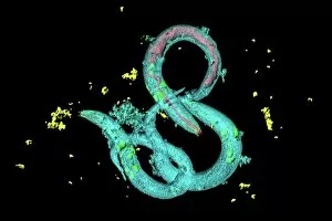
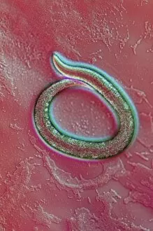
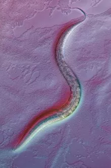
"Unveiling the Secrets of Life: Exploring Model Organisms Through Microscopy" Witness the intricate world of model organisms through a light micrograph, as C


For sale as Licensed Images
Choose your image, Select your licence and Download the media
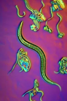
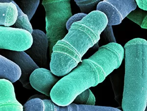
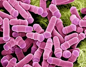
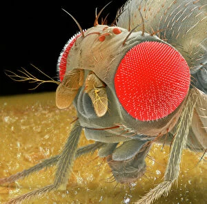
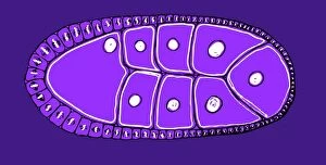
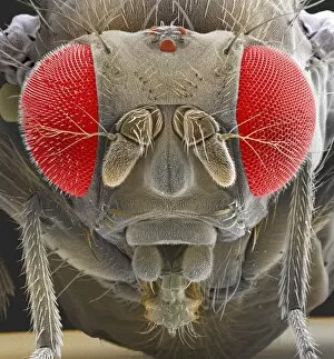
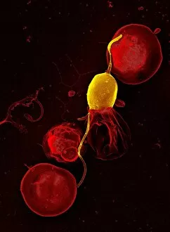
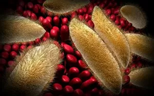
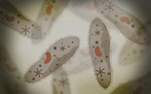
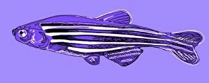
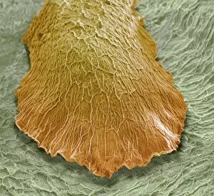
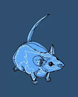
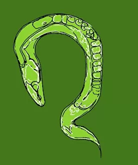
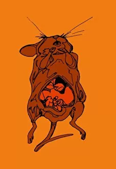
"Unveiling the Secrets of Life: Exploring Model Organisms Through Microscopy" Witness the intricate world of model organisms through a light micrograph, as C. Elegans worms gracefully navigate their environment. Delve into the fascinating realm of cell division with dividing yeast cells captured in stunning detail by scanning electron microscopy (SEM). Marvel at another glimpse of C. Elegans worms under a light micrograph, revealing their unique features and behaviors. Explore the microscopic universe further with SEM imagery showcasing dividing yeast cells, offering insights into their complex reproductive processes. Discover the captivating beauty of fruit flies through SEM Z340 / 0699, unveiling their delicate structures and intricate patterns. Uncover an astonishing view of a mouse malaria parasite via SEM, shedding light on its morphology and potential targets for intervention. Immerse yourself in the enchanting world of fruit flies once again with SEM Z340 / 0700, capturing their ethereal essence from a different perspective. Picture No. 12479375 takes you on an extraordinary journey into unknown realms where unseen wonders await your exploration. Peer closely at a mesmerizing microscopic view of paramecium – an organism that holds secrets to understanding cellular dynamics and evolution. Embark on a conceptual voyage as you encounter Euglena's abstract image – representing both simplicity and complexity within this remarkable model organism's existence. Explore yet another conceptual image depicting paramecium - inviting contemplation about its role in scientific research and our understanding of life itself.