Molecular Biology Collection (#22)

"Molecular Biology: Unveiling the Secrets of Life at a Microscopic Level" Amitriptyline Antidepressant Molecule: Exploring the Chemical Pathways to Mental Well-being

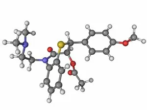
Background imageMolecular Biology Collection: Diltiazem molecule

Diltiazem molecule
Diltiazem, molecular model. This calcium channel blocker drug is used to treat hypertension (high blood pressure) and angina

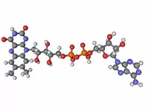
Background imageMolecular Biology Collection: Flavin adenine dinucleotide molecule

Flavin adenine dinucleotide molecule
Flavin adenine dinucleotide, molecular model. This coenzyme is an important intermediary in biological oxidations and reductions

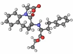
Background imageMolecular Biology Collection: Benazepril high blood pressure drug

Benazepril high blood pressure drug
Benazepril, molecular model. This hypertension (high blood pressure) drug is marketed as Lotensin. Atoms are represented as spheres and are colour-coded: carbon (grey), hydrogen (white)

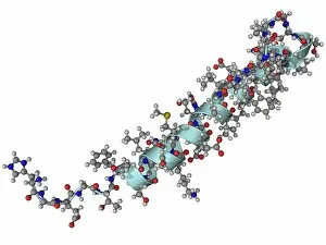
Background imageMolecular Biology Collection: Exendin-4 diabetes drug molecule

Exendin-4 diabetes drug molecule
Exendin-4, molecular model. This drug, which increases insulin release in those with type 2 diabetes, is extracted from the saliva of the Gila monster (Heloderma suspectum) lizard

Background imageMolecular Biology Collection: Artemisinin malaria drug molecule

Artemisinin malaria drug molecule
Artemisinin, molecular model. This drug, which is used to treat malaria, is extracted from the leaves of the wormwood plant (Artemisia annua)

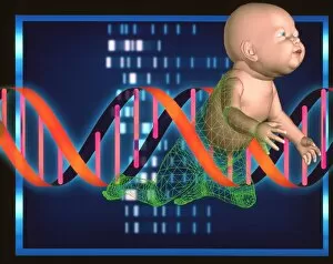
Background imageMolecular Biology Collection: Designer baby

Designer baby. Conceptual computer artwork of a human baby, a DNA (deoxyribonucleic acid) double helix (centre) and genetic sequences (white bands), representing a designer baby

Background imageMolecular Biology Collection: DNA, symbolic sculpture

DNA, symbolic sculpture
Conceptual 3d DNA-like sculpture depicting the genetic code that is unique for every individual. DNA controls the development and functioning of every living organism

Background imageMolecular Biology Collection: DNA

DNA. Conceptual computer artwork of the double helix DNA (deoxyribonucleic acid) molecule (lower centre), a female face (centre right) and a DNA autoradiogram (upper centre)

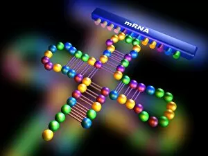
Background imageMolecular Biology Collection: Genetic translation

Genetic translation, computer diagram. This process uses genetic information to direct the synthesis of proteins. The main molecules involved are two types of RNA (ribonucleic acid)

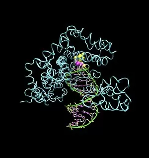
Background imageMolecular Biology Collection: Taq polymerase replicating DNA

Taq polymerase replicating DNA, molecular model. The Taq polymerase is blue, the two strands of DNA are green, and the nucleotide bases are grey (carbon), pink (nitrogen)

Background imageMolecular Biology Collection: Topoisomerase 1 / DNA complex

Topoisomerase 1 / DNA complex
Human topoisomerase 1/DNA complex, molecular model. Topoisomerase 1 is an enzyme that assists in uncoiling DNA. DNA (deoxyribonucleic acid) is usually stored in a supercoiled form

Background imageMolecular Biology Collection: Computer artwork of part of a beta DNA molecule

Computer artwork of part of a beta DNA molecule
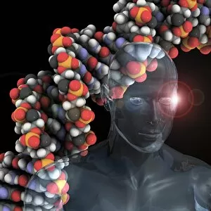
DNA molecule. Computer representation of a segment of the molecule Deoxyribonucleic Acid (DNA). This structure contains all the inherited instructions necessary for the development of a living organ
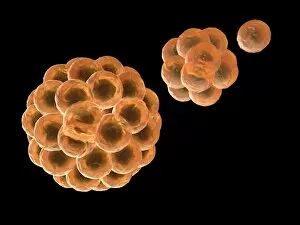
Background imageMolecular Biology Collection: Stem cells division, computer artwork

Stem cells division, computer artwork
Computer artwork depicting division of stem cells. A stem cell is an undifferentiated call that can divide indefinitely in culture

Background imageMolecular Biology Collection: DNA

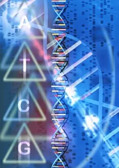
DNA

Background imageMolecular Biology Collection: Lunar DNA library

Lunar DNA library, conceptual computer artwork. The surface of the Moon is across bottom, with the Earth in the background

Background imageMolecular Biology Collection: Gene activator protein

Gene activator protein. Computer artwork of catabolite gene activator protein (CAP, blue and red) bound to a molecule of deoxyribonucleic acid (DNA)

Background imageMolecular Biology Collection: Cryptochrome and rhodopsin pigments

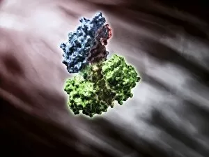
Cryptochrome and rhodopsin pigments. Rhodopsin is a pigment found in the rod photoreceptor cells in the retina of the human eye, where it absorbs light falling on the retina

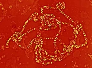
Background imageMolecular Biology Collection: Giant chromosomes, light micrograph

Giant chromosomes, light micrograph. These giant (polytene) chromosomes are from the salivary glands of the fruit fly Drosophila melanogaster

Background imageMolecular Biology Collection: Cholera toxin, artwork

Cholera toxin, artwork
Cholera toxin, molecular structure. Cholera is an infectious intestinal disease caused by this toxin produced by the Gram-negative bacterium Vibrio cholerae

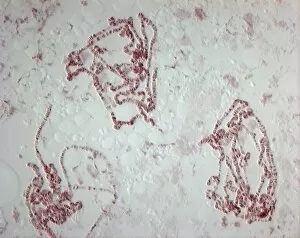
Background imageMolecular Biology Collection: Giant chromosome, light micrograph

Giant chromosome, light micrograph

Background imageMolecular Biology Collection: Physalis mottle virus capsid protein

Physalis mottle virus capsid protein, molecular model

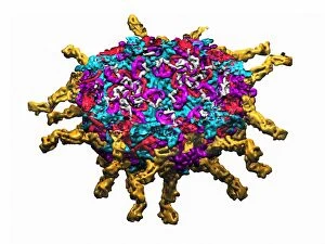
Background imageMolecular Biology Collection: Bovine rotavirus capsid protein

Bovine rotavirus capsid protein, molecular model

Background imageMolecular Biology Collection: Reovirus particle, molecular model

Reovirus particle, molecular model

Background imageMolecular Biology Collection: Nudaurelia capensis omega virus

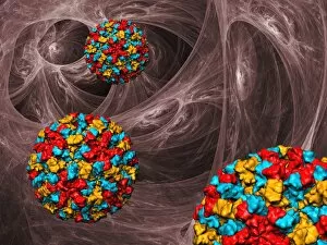
Nudaurelia capensis omega virus. Computer model of the capsid of the Nudaurelia capensis omega virus

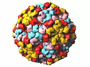
Background imageMolecular Biology Collection: Poliovirus particle, molecular model

Poliovirus particle, molecular model
Poliovirus virus particle, computer model

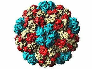
Background imageMolecular Biology Collection: Coxsackie B3 virus particle

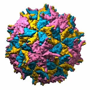
Coxsackie B3 virus particle. Computer artwork of the capsid of the Coxsackie B3 virus, with proteins represented by coloured blobs. The capsid is icosahedral in shape, with 20 triangular faces

Background imageMolecular Biology Collection: Brome grass mosaic virus particle

Brome grass mosaic virus particle
Brome grass mosaic virus. Computer model of the capsid of the Brome grass mosaic virus

Background imageMolecular Biology Collection: Norwalk virus particles, artwork

Norwalk virus particles, artwork
Norwalk viruses. Computer artwork of Norwalk virus particles

Background imageMolecular Biology Collection: Simian virus 40 particle, molecular model

Simian virus 40 particle, molecular model
Simian virus 40. Computer model of the capsid of the Simian virus 40 virion

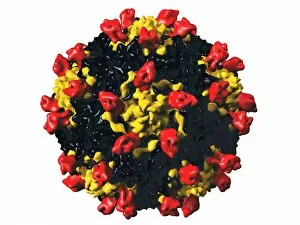
Background imageMolecular Biology Collection: Nodamura virus particle, molecular model

Nodamura virus particle, molecular model
Nodamura virus. Computer model of the capsid of the Nodamura virus

Background imageMolecular Biology Collection: Human genome, conceptual artwork

Human genome, conceptual artwork
Human genome, conceptual computer artwork

Background imageMolecular Biology Collection: Bacteriophage alpha 3, artwork

Bacteriophage alpha 3, artwork
Bacteriophage alpha 3. Computer model showing the structure of the protein capsid of bacteriophage alpha 3. Bacteriophages are viruses that infect bacteria

Background imageMolecular Biology Collection: Outer membrane receptor protein molecule

Outer membrane receptor protein molecule
Outer membrane receptor protein. Molecular model of FecA an outer membrane receptor protein

Background imageMolecular Biology Collection: Viral transfer of bacterial DNA, artwork

Viral transfer of bacterial DNA, artwork
Viral transfer of bacterial DNA. Computer artwork showing the process of transduction, whereby genetic information is transferred from one bacterium (right column) to another (left column)

Background imageMolecular Biology Collection: Heterogeneous nuclear ribonucleoprotein

Heterogeneous nuclear ribonucleoprotein. Molecular model of a heterogeneous nuclear ribonucleoprotein (hnRNP, orange) complexed with an RNA (ribonucleic acid, yellow) molecule

Background imageMolecular Biology Collection: PPAR regulatory molecule

PPAR regulatory molecule. Molecular model of peroxisome proliferator-activated receptor gamma (PPARG, pink) bound to a DNA (deoxyribonucleic acid) molecule (orange and green)

Background imageMolecular Biology Collection: Plant cell wall, diagram

Plant cell wall, diagram

Background imageMolecular Biology Collection: Gene splicing, diagram

Gene splicing, diagram
Gene splicing. Diagram showing eight stages involved in gene splicing by a complex known as a spliceosome. The process (top) involves removing a fragment known as an intron (orange)

Background imageMolecular Biology Collection: HIV replication cycle, diagram

HIV replication cycle, diagram
HIV replication cycle. Diagram and magnified views showing the retroviral process by which the human immunodeficiency virus (HIV) replicates

Background imageMolecular Biology Collection: Chromatin condensation, diagram

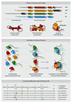
Chromatin condensation, diagram. This sequence, from right to left, shows the stages by which a long strand of DNA (deoxyribonucleic acid)

Background imageMolecular Biology Collection: Genetic molecular mechanisms, artwork

Genetic molecular mechanisms, artwork

Background imageMolecular Biology Collection: Protein translation, artwork

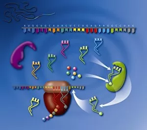
Protein translation, artwork
Protein translation. Artwork showing the process of translation, the final stage of the production of proteins from the genetic code

Background imageMolecular Biology Collection: Transcription initiation complex, diagram

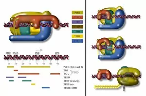
Transcription initiation complex, diagram
Transcription initiation complex. Diagram of the complex formed by the molecules involved in the initiation of transcription

Background imageMolecular Biology Collection: Cytoskeleton components, artwork

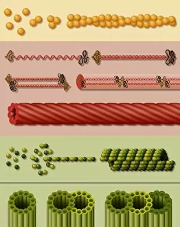
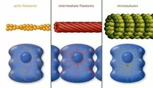
Cytoskeleton components, artwork. Three main components of the cytoskeleton, the internal support structure of a cell, are shown here

Background imageMolecular Biology Collection: DNA replication fork, artwork

DNA replication fork, artwork
DNA replication fork. Diagram showing the cyclic sequence (right) for replication of DNA (deoxyribonucleic acid). Details of the DNA are at left

Background imageMolecular Biology Collection: Eukaryotic flagellum structure, artwork

Eukaryotic flagellum structure, artwork
Eukaryotic flagellum structure. Cutaway artwork showing the internal structure of the flagellum in eukaryotic cells. Flagella are tail-like projections used for cell locomotion

Background imageMolecular Biology Collection: Cytoskeleton components, diagram

Cytoskeleton components, diagram. The cytoskeleton is the internal support structure of a cell, composed of filaments of various diameters in nanometres (nm)

Background imageMolecular Biology Collection: Histone structures, diagram

Histone structures, diagram. Histone cores are cylindrical structures around which the genetic molecule DNA (deoxyribonucleic acid) is wound with other proteins to form chromatin


For sale as Licensed Images

Choose your image, Select your licence and Download the media

"Molecular Biology: Unveiling the Secrets of Life at a Microscopic Level" Amitriptyline Antidepressant Molecule: Exploring the Chemical Pathways to Mental Well-being. Watson and Crick, DNA Discovers: Pioneers Who Unraveled the Double Helix Structure, Revolutionizing Genetics. Creatine Amino Acid Molecule: Fueling Muscles and Unlocking Athletic Performance Potential. Microscopic View of Human Respiratory Syncytial Virus: Understanding the Culprit Behind Common Respiratory Infections. Embryo Development 24-36 Hours After Fertilization: Witnessing the Miracle of Life from Conception to Early Growth. Zinc Fingers Bound to a DNA Strand: Decoding Genetic Instructions for Cellular Functions and Disease Prevention. SARS Coronavirus Protein: Investigating Viral Mechanisms for Effective Control and Treatment Strategies. Vioxx Drug Molecule: Analyzing Medications' Molecular Structures for Enhanced Safety and Efficacy in Pain Management. Levofloxacin Antibiotic Molecule: Combating Bacterial Infections with Precision-targeted Therapies. Electrophoresis of RNA: Separating Genetic Material to Study Gene Expression Patterns in Health and Disease Mitochondrial DNA - The Powerhouses Within Our Cells Holding Clues to Evolutionary History Cone Snail Venom Component Molecule.