Molecules Collection (#10)

"Molecules

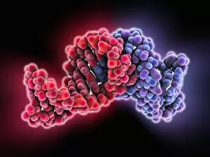
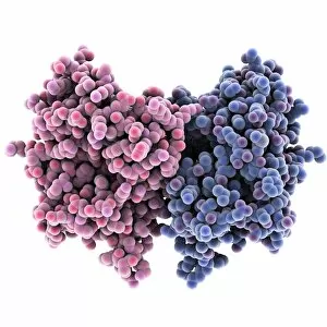
Background imageMolecules Collection: Bacterial RNA plasmid loop-loop complex

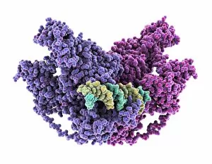
Bacterial RNA plasmid loop-loop complex, molecular model. This strand of ribonucleic acid (RNA) is part of a plasmid, the loop of genetic material found in bacterial cells
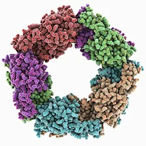
Background imageMolecules Collection: Cat allergen protein, molecular model

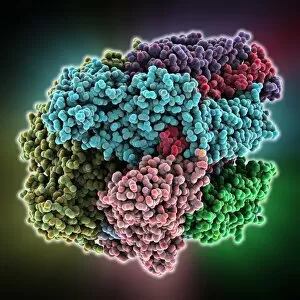
Cat allergen protein, molecular model
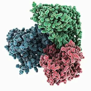
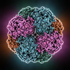
Cat allergen protein. Molecular model of the tetrameric form of the major cat allergen fel d 1 (Felis domesticus allergen 1)

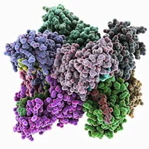
Background imageMolecules Collection: Methane monooxygenase enzyme

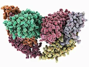
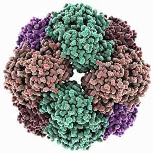
Methane monooxygenase enzyme, molecular model. This is the particulate methane monooxygenase (pMMO) form of this metalloenzyme, an integral membrane protein that contains copper and zinc

Background imageMolecules Collection: Atomic interactions, conceptual image C013 / 5595

Atomic interactions, conceptual image C013 / 5595
Atomic interactions, conceptual image. Computer artwork representing the interactions between atomic and sub-atomic particles

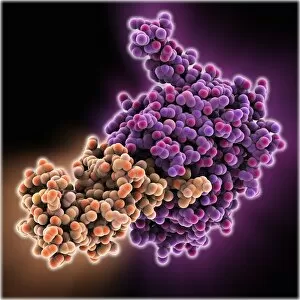
Background imageMolecules Collection: FP2 malaria protease enzyme complex

FP2 malaria protease enzyme complex, molecular model. This complex consists of the falcipain-2 (FP2) protease enzyme (purple, right) bound to a cystatin (orange, left), a form of protease inhibitor

Background imageMolecules Collection: Follicle-stimulating hormone complex C015 / 0945

Follicle-stimulating hormone complex C015 / 0945
Follicle-stimulating hormone (FSH) complex with receptor, molecular model. FSH helps to regulate human sexual development and reproductive processes. In females, it acts on follicles in the ovaries

Background imageMolecules Collection: Follicle-stimulating hormone complex C015 / 0944

Follicle-stimulating hormone complex C015 / 0944
Follicle-stimulating hormone (FSH) complex with receptor, molecular model. FSH helps to regulate human sexual development and reproductive processes. In females, it acts on follicles in the ovaries

Background imageMolecules Collection: Glutamate transporter protein

Glutamate transporter protein, molecular model. This is a membrane protein that facilitates the uptake of glutamate by a cell, thus playing an important role in neurology in higher organisms

Background imageMolecules Collection: Purple bacterium photosynthesis centre

Purple bacterium photosynthesis centre, molecular model. Purple bacteria are phototrophic bacteria that produce energy through photosynthesis

Background imageMolecules Collection: Oestrogen related receptor-DNA complex

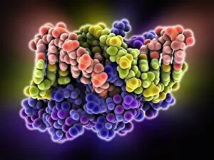
Oestrogen related receptor-DNA complex. Molecular model of human estrogen related receptor-2 (heRR-2, purple) binding to a strand of DNA (deoxyribonucleic acid, red and yellow-green)

Background imageMolecules Collection: Lambda repressor-operator complex

Lambda repressor-operator complex. Molecular model of the lambda repressor protein (red and green) binding to a region of DNA (deoxyribonucleic acid, orange and blue) known as the lambda operator

Background imageMolecules Collection: Repair protein and DNA, molecular model

Repair protein and DNA, molecular model
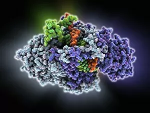
Repair protein and DNA. Molecular model of the Ku heterodimer (grey, blue and purple) bound to a strand of DNA (deoxyribonucleic acid, orange and green) as part of the repair process

Background imageMolecules Collection: Max transcription factor-DNA complex

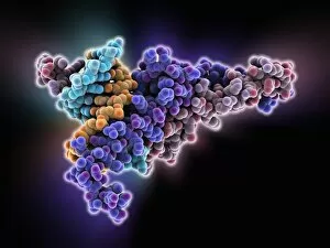
Max transcription factor-DNA complex. Molecular model of the Max transcription factor (purple and red) bound to a strand of DNA (deoxyribonucleic acid, light blue and orange)

Background imageMolecules Collection: Antibody fragment-lysozyme complex

Antibody fragment-lysozyme complex
fab d1.3, , chicken egg white lysozyme, protein, biomolecule, macromolecule, lysozyme, enzyme, fab, antigen, antibody, biochemistry, biology, molecular biology, proteomics, artwork, illustration

Background imageMolecules Collection: Bacterial regulator-DNA complex

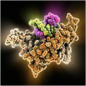
Bacterial regulator-DNA complex. Molecular model of a complex formed between a bacterial regulator called SarA (orange and brown) and a fragment of DNA (pink and yellow-green strands)

Background imageMolecules Collection: Bacterial protein-chaperone complex

Bacterial protein-chaperone complex. Molecular model of a bacterial effector protein binding to a chaperone protein that helps prevent keep the bacterial protein in an unfolded or partially folded

Background imageMolecules Collection: Hin recombinase-DNA complex

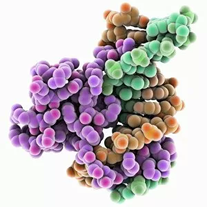
Hin recombinase-DNA complex. Molecular model of the Hin recombinase protein (pink) bound to a double helix (green and orange) strand of DNA (deoxyribonucleic acid)

Background imageMolecules Collection: Tumour suppressor protein molecular model C016 / 2065

Tumour suppressor protein molecular model C016 / 2065
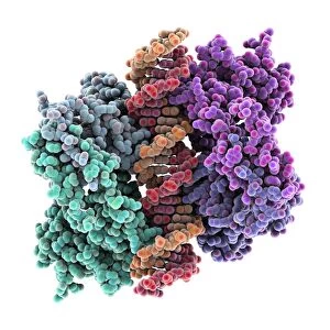
Tumour suppressor protein. Molecular model of the tumour suppressor protein p53 (left and right) bound to a molecule of DNA (deoxyribonucleic acid, down centre) at the p53 response element

Background imageMolecules Collection: Restriction enzyme cutting DNA

Restriction enzyme cutting DNA
Fragment of DNA bound by the restriction endonucleaseEcoRI. The protein is a dimer, with each subunitable to bind and cut one strand of DNA

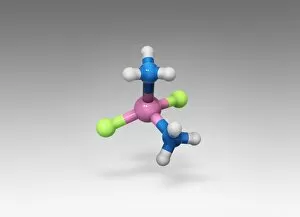
Background imageMolecules Collection: Cisplatin drug molecule

Cisplatin drug molecule. Computer artwork showing the structure of a molecule of the chemotherapy drug Cisplatin. This drug is used to treat a number of cancers, including sarcomas

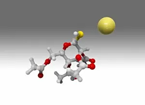
Background imageMolecules Collection: Auranofin drug molecule

Auranofin drug molecule. Computer artwork showing the structure of a molecule of the drug auranofin. Auranofin, marketed under the name Ridaura

Background imageMolecules Collection: Poly(A)-binding protein and RNA complex

Poly(A)-binding protein and RNA complex. Computer model showing the structure of a poly(A)-binding protein (PABP) molecule bound to the poly(A)

Background imageMolecules Collection: Vitamin B1 molecule

Vitamin B1 molecule. Computer model showing the structure of a molecule of vitamin B1 (thiamine). Vitamin B1 is an essential nutrient that humans are unable to produce

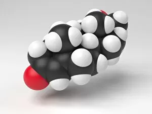
Background imageMolecules Collection: Testosterone hormone molecule

Testosterone hormone molecule. Computer model showing the structure of a molecule of the male sex hormone testosterone. Testosterone is the main human androgen

Background imageMolecules Collection: Progesterone hormone molecule

Progesterone hormone molecule. Computer model showing the structure of a molecule of the hormone progesterone. Progesterone is produced in the ovaries of women and the testes of men

Background imageMolecules Collection: Glyphosate weed killer molecule

Glyphosate weed killer molecule. Computer model showing the molecular structure of a molecule of the herbicide glyphosate. Glyphosate is a widely used herbicide

Background imageMolecules Collection: Ibuprofen molecule

Ibuprofen molecule. Computer artwork showing the structure of a molecule of the painkilling (analgesic) drug ibuprofen. Ibuprofen is a non-steroidal anti-inflammatory drug (NSAID)

Background imageMolecules Collection: Thrombin protein, molecular model C015 / 7074

Thrombin protein, molecular model C015 / 7074
Thrombin protein, molecular model. Thrombin is an enzyme involved in the blood coagulation (clotting) process. It converts fibrinogen (a soluble plasma glycoprotein synthesised in the liver)

Background imageMolecules Collection: Simian virus (SV40) large T antigen C015 / 7070

Simian virus (SV40) large T antigen C015 / 7070
Simian virus (SV40) large T antigen, molecular model. This antigen is from the simian vacuolating virus 40 (SV40). Large T antigens play a role in regulating the viral life cycle of

Background imageMolecules Collection: Bacterial alpha-hemolysin toxin C015 / 7067

Bacterial alpha-hemolysin toxin C015 / 7067
Bacterial alpha-hemolysin toxin, molecular model. This toxin is secreted by the bacterium Staphylococcus aureus. It is an example of a pore-forming toxin

Background imageMolecules Collection: Sir3 gene silencer acting on DNA C015 / 7062

Sir3 gene silencer acting on DNA C015 / 7062
Sir3 gene silencer acting on DNA, molecular model. Sir3 (purple and grey) is acting on a circular strand of DNA (deoxyribonucleic acid, red)

Background imageMolecules Collection: Sir3 gene silencer acting on DNA C015 / 7061

Sir3 gene silencer acting on DNA C015 / 7061
Sir3 gene silencer acting on DNA

Background imageMolecules Collection: TFAM transcription factor bound to DNA C015 / 7060

TFAM transcription factor bound to DNA C015 / 7060
TFAM transcription factor bound to DNA, molecular model. Human mitochondrial transcription factor A (TFAM, pink) bound to a strand of DNA (deoxyribonucleic acid, orange and green)

Background imageMolecules Collection: Caffeine molecule

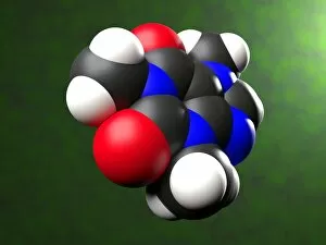
Caffeine molecule. Computer artwork showing the structure of a molecule of the alkaloid stimulant and legal drug caffeine. Caffeine is found in drinks such as tea, coffee, and fizzy drinks

Background imageMolecules Collection: Serotonin molecule

Serotonin molecule. Computer model showing the structure of a molecule of the neurotransmitter (nerve signalling chemical) serotonin (5-hydroxytryptamine)

Background imageMolecules Collection: Aspirin molecule

Aspirin molecule. Computer artwork showing the structure of a molecule of aspirin (acetylsalicylic acid). Atoms are represented as spheres and are colour-coded: carbon (black), hydrogen (white)

Background imageMolecules Collection: Aspirin in action

Aspirin in action. Computer artwork showing how aspirin has its effect. Aspirin (acetylsalicylic acid) is converted to salicylic acid and acetic acid in the body

Background imageMolecules Collection: Peroxiredoxin 4 antioxidant enzyme C015 / 7022

Peroxiredoxin 4 antioxidant enzyme C015 / 7022
Peroxiredoxin 4 antioxidant enzyme, molecular model. This enzyme, also called peroxiredoxin IV (PrxIV), plays a catalytic role in cell metabolism on the endoplasmic reticulum

Background imageMolecules Collection: Mandelate racemase enzyme C015 / 7021

Mandelate racemase enzyme C015 / 7021
Mandelate racemase enzyme, molecular model. This bacterial enzyme is an example of a muconate lactonizing enzyme, and plays a key role in the metabolism of soil and aquatic bacteria

Background imageMolecules Collection: Mandelate racemase enzyme C015 / 7020

Mandelate racemase enzyme C015 / 7020
Mandelate racemase enzyme, molecular model. This bacterial enzyme is an example of a muconate lactonizing enzyme, and plays a key role in the metabolism of soil and aquatic bacteria

Background imageMolecules Collection: Restriction enzyme and DNA C015 / 6941

Restriction enzyme and DNA C015 / 6941
Restriction enzyme and DNA. Molecular model showing an EcoRI endonuclease enzyme (purple) bound to a DNA (deoxyribonucleic acid) strand (blue). EcoRI is an enzyme isolated from strains of E

Background imageMolecules Collection: Human serum albumin molecule C015 / 6938

Human serum albumin molecule C015 / 6938
Human serum albumin, molecular model. Albumin is the most abundant protein in human blood plasma. It is seen here complexed with stearic acid, a saturated fatty acid

Background imageMolecules Collection: Nucleoplasmin histone-chaperone protein C015 / 6915

Nucleoplasmin histone-chaperone protein C015 / 6915
Nucleoplasmin histone-chaperone protein, molecular model. This is nucleoplasmin 2 (Npm2), a chaperone that acts on human nucleoplasmin. Chaparone proteins play a key role in aiding protein folding

Background imageMolecules Collection: Methyltransferase complexed with DNA C016 / 2033

Methyltransferase complexed with DNA C016 / 2033
Methyltransferase complexed with DNA, molecular model. The strand of DNA (deoxyribonucleic acid, green and blue) is enclosed by DNA methyltransferase 1 (DNMT-1, green and pink)

Background imageMolecules Collection: Newcastle disease virus protein C015 / 6912

Newcastle disease virus protein C015 / 6912
Newcastle disease virus protein, molecular model. This is a hemagglutinin-neuromidase (HN) protein that enables the virus to bind to and infect host cells

Background imageMolecules Collection: Methyltransferase complexed with DNA C016 / 2032

Methyltransferase complexed with DNA C016 / 2032
Methyltransferase complexed with DNA, molecular model. The strand of DNA (deoxyribonucleic acid, green and yellow) is enclosed by DNA methyltransferase 1 (DNMT-1, purple and pink)

Background imageMolecules Collection: NpmA methyltransferase C016 / 2031

NpmA methyltransferase C016 / 2031
NpmA methyltransferase, molecular model. Methyltransferase enzymes act to add methyl groups to nucleic acids such as DNA, a process called DNA methylation

Background imageMolecules Collection: NpmA methyltransferase C016 / 2030

NpmA methyltransferase C016 / 2030
NpmA methyltransferase, molecular model. Methyltransferase enzymes act to add methyl groups to nucleic acids such as DNA, a process called DNA methylation


For sale as Licensed Images

Choose your image, Select your licence and Download the media

"Molecules: The Building Blocks of Life and Beyond" From the intricate workings of an anaesthetic inhibiting an ion channel C015/6718 to the genius mind of James Clerk Maxwell, they have captivated scientists and artists alike. With their diverse structures and functions, they hold the key to understanding life at its core. Delving into the world of proteins, we witness their secondary structure through mesmerizing artwork that unveils their complexity. Meanwhile, the caffeine drug molecule keeps us awake while bacterial ribosomes tirelessly synthesize proteins within our cells. Vitamin B12's molecular model reminds us of nature's intricate design as zinc fingers elegantly bind to a DNA strand, orchestrating genetic processes. And who can forget capsaicin - the fiery molecule responsible for giving chili peppers their spicy kick? But molecules aren't limited to just earthly matters; they extend beyond our planet's boundaries. Oxytocin neurotransmitter molecules remind us of love's chemical connection while praziquantel parasite drugs combat infections in distant lands. Interferon molecules stand tall as defenders against viral invasions, showcasing our body's remarkable defense mechanisms. And amidst all this scientific wonder lies a breathtaking sight - Aurora Borealis dancing over a snow-covered coniferous forest in Northern Finland. Intricate and awe-inspiring, these glimpses into the molecular world remind us that there is so much more than meets the eye. From unlocking medical breakthroughs to unraveling nature's mysteries or simply marveling at captivating artistry – they can truly extraordinary entities shaping our understanding of life itself.