
Musculoskeletal System Collection (#2)
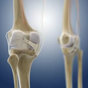
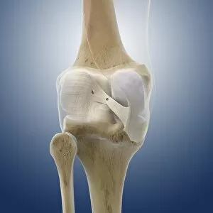
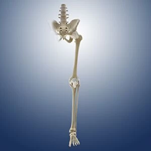
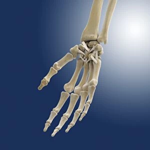
The musculoskeletal system is a complex network of bones, muscles, ligaments, and nerves that work together to provide support and movement for the body

For sale as Licensed Images
Choose your image, Select your licence and Download the media
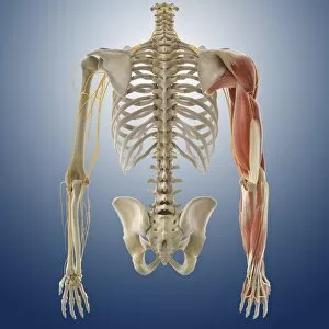
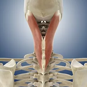
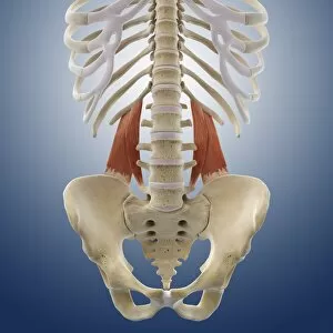
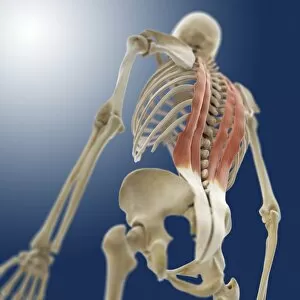
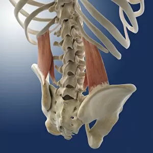
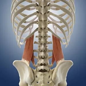
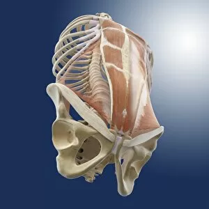
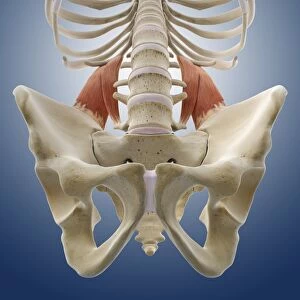
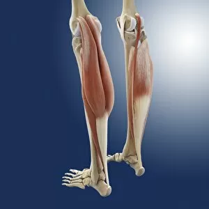
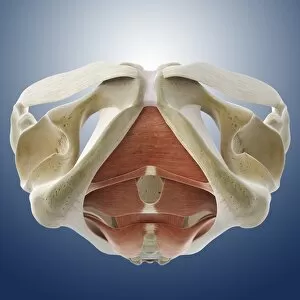
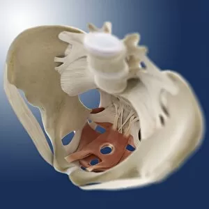
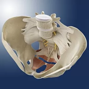
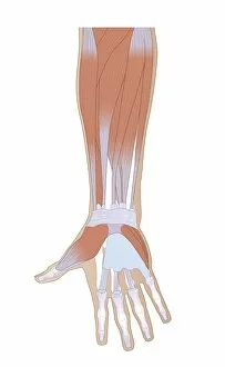
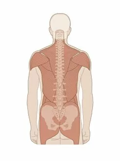
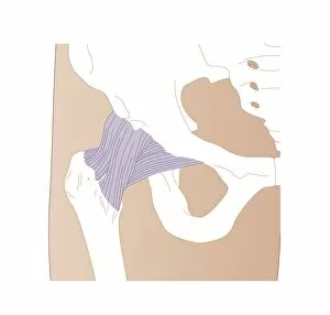
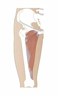
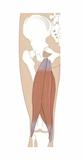
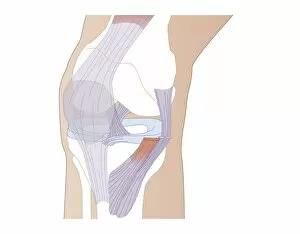
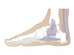
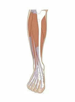
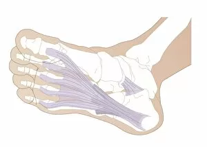
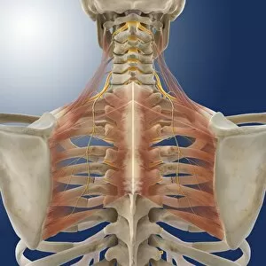
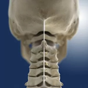
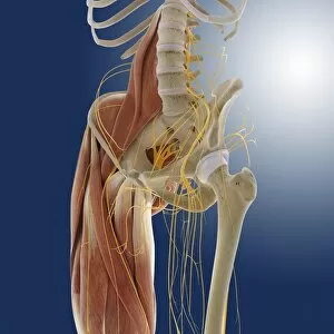
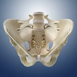
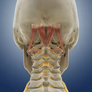
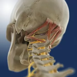
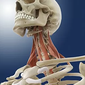
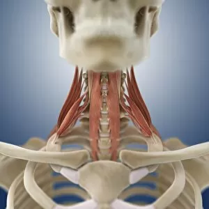
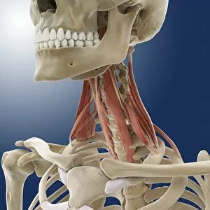
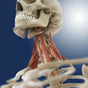
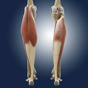
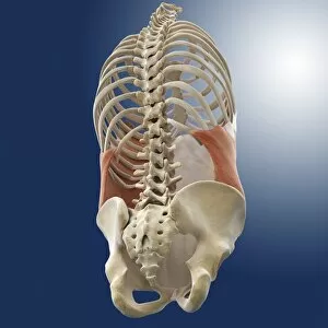
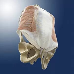
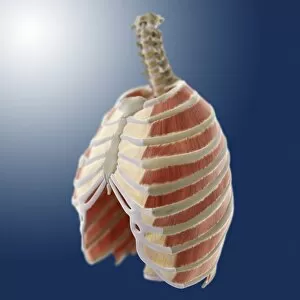
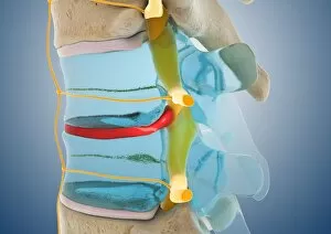
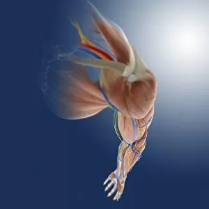
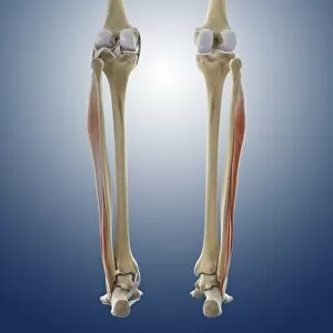
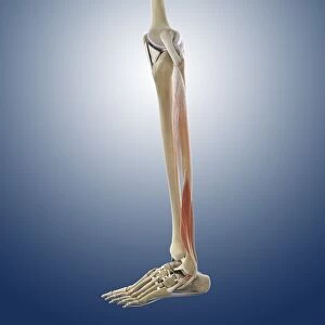
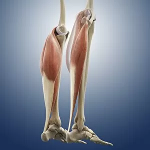
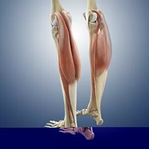
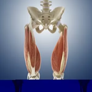
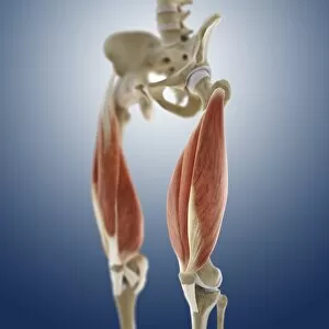
The musculoskeletal system is a complex network of bones, muscles, ligaments, and nerves that work together to provide support and movement for the body. From the outer ankle ligaments to the inner ankle ligaments, every part plays a crucial role in maintaining stability and flexibility. Artwork C013 / 4452 beautifully depicts the intricate structure of the outer ankle ligaments, while artwork C013 / 4451 showcases the delicate nature of the inner ankle ligaments. These illustrations serve as a visual reminder of how these ligaments enable us to walk, run, and perform various activities with ease. Moving up towards the female muscular system artwork, we witness the strength and power that lies within our muscles. The human upper body illustration further emphasizes this by showcasing not only bones but also muscles and even our circulatory system. It reminds us that without proper muscle function, our bodies would lack mobility and vitality. Delving deeper into specific areas of our body's anatomy, we come across artwork depicting neck muscles and nerves (artwork), upper arm muscles (artwork), phrenic nerves alongside diaphragm (artwork), suboccipital muscles along with nerve (artwork C014 / 5097). These illustrations highlight how each muscle group works in harmony with its corresponding nerve supply to facilitate movement or maintain posture. Artworks such as arm anatomy (C013 / 4583) showcase detailed structures like tendons connecting muscle to bone while buttock muscles (C013 / 4414) remind us that even seemingly insignificant areas play vital roles in everyday movements like sitting or standing. Lastly, a cross-section biomedical illustration provides an overall view of an adult male's musculoskeletal system. This comprehensive representation serves as a reminder that all these individual parts are interconnected - from bones providing structural support to muscles enabling motion - creating an intricate web necessary for optimal functioning.