Musculoskeletal System Collection (#4)

The musculoskeletal system is a complex network of bones, muscles, ligaments, and nerves that work together to provide support and movement for the body

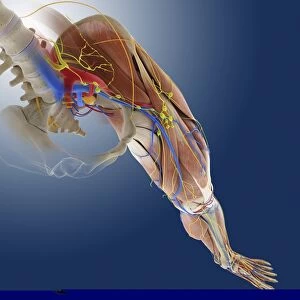
Background imageMusculoskeletal System Collection: Leg anatomy, artwork C013 / 4493

Leg anatomy, artwork C013 / 4493
Leg anatomy. Computer artwork of an oblique anterior view looking down a human leg, showing muscles and tendons (brown and white), bones and ligaments (white), arteries and veins (red and blue)

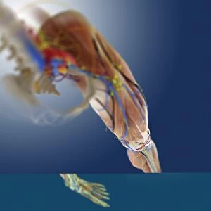
Background imageMusculoskeletal System Collection: Leg anatomy, artwork C013 / 4494

Leg anatomy, artwork C013 / 4494
Leg anatomy. Computer artwork of an oblique anterior view looking down a human leg, showing muscles and tendons (brown and white), bones and ligaments (white), arteries and veins (red and blue)

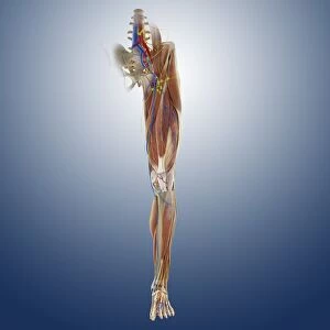
Background imageMusculoskeletal System Collection: Leg ligaments, artwork

Leg ligaments, artwork
Leg anatomy. Computer artwork of an anterior view of a human leg, showing the muscles and tendons (brown and white), bones and ligaments (white), arteries and veins (red and blue), nerves (yellow)

Background imageMusculoskeletal System Collection: Foot muscles, artwork C013 / 4487

Foot muscles, artwork C013 / 4487
Foot muscles. Computer artwork of a view looking down on some of the muscles and tendons (red and white) of the upper surface of the foot

Background imageMusculoskeletal System Collection: Foot muscles, artwork C013 / 4486

Foot muscles, artwork C013 / 4486
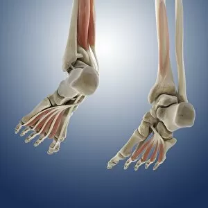
Foot muscles. Computer artwork of an oblique anterior view of some of the muscles and tendons (red and white) of the upper surface of the foot

Background imageMusculoskeletal System Collection: Foot muscles, artwork C013 / 4485

Foot muscles, artwork C013 / 4485
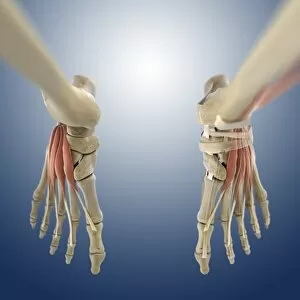
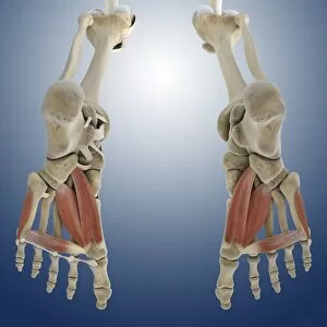
Foot muscles. Computer artwork of a view from below of some of the muscles and tendons (red and white) of the sole of the foot

Background imageMusculoskeletal System Collection: Foot muscles, artwork C013 / 4483

Foot muscles, artwork C013 / 4483
Foot muscles. Computer artwork of an oblique view from below and behind of some of the muscles and tendons (red and white) of the sole of the foot

Background imageMusculoskeletal System Collection: Foot muscles, artwork C013 / 4478

Foot muscles, artwork C013 / 4478
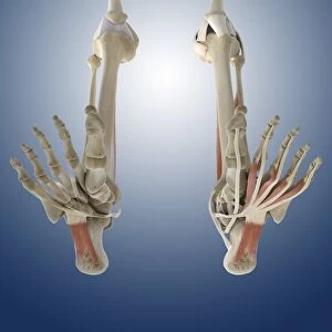
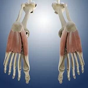
Foot muscles. Computer artwork of a view from below of some of the muscles and tendons (red and white) of the sole of the foot

Background imageMusculoskeletal System Collection: Foot muscles, artwork C013 / 4477

Foot muscles, artwork C013 / 4477
Foot muscles. Computer artwork of a view from below of some of the muscles and tendons (red and white) of the sole of the foot

Background imageMusculoskeletal System Collection: Outer ankle ligaments, artwork C013 / 4456

Outer ankle ligaments, artwork C013 / 4456
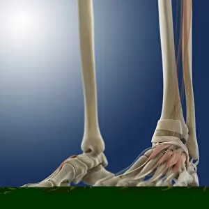
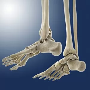
Outer ankle ligaments. Computer artwork of the bones and ligaments (white) of the feet and ankles from an oblique side view, with the outer side of the right foot at left

Background imageMusculoskeletal System Collection: Outer ankle ligaments, artwork C013 / 4455

Outer ankle ligaments, artwork C013 / 4455
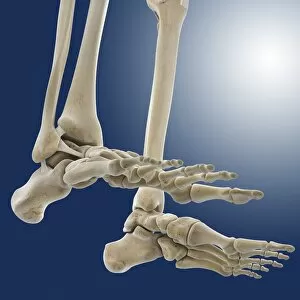
Outer ankle ligaments. Computer artwork of the bones and ligaments (white) of the feet and ankles from an oblique side view, with the outer side of the left foot at upper centre

Background imageMusculoskeletal System Collection: Hip ligaments, artwork C013 / 4434

Hip ligaments, artwork C013 / 4434
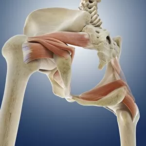
Hip ligaments. Computer artwork of a posterior oblique view of the hip bones and ligaments (white). At upper right the lumbar spine ends in the sacrum and coccyx (tailbone)

Background imageMusculoskeletal System Collection: Hip ligaments, artwork C013 / 4433

Hip ligaments, artwork C013 / 4433
Hip ligaments. Computer artwork of a posterior view of the hip bones and ligaments (white). At centre the lumbar spine ends in the sacrum and coccyx (tailbone)

Background imageMusculoskeletal System Collection: Hip ligaments, artwork C013 / 4432

Hip ligaments, artwork C013 / 4432
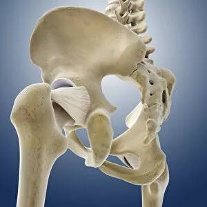
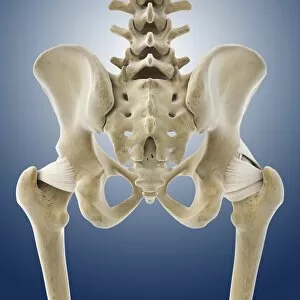
Hip ligaments. Computer artwork of an anterior view of the hip bones and ligaments (white). At centre the lumbar spine ends in the sacrum and coccyx (tailbone)

Background imageMusculoskeletal System Collection: Hip anatomy, artwork C013 / 4430

Hip anatomy, artwork C013 / 4430
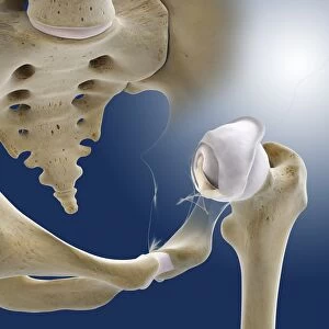
Hip anatomy. Computer artwork of the head of the left femur (centre right) articulating with the left-hand side of the pelvis to form the hip joint

Background imageMusculoskeletal System Collection: Leg muscles, artwork C013 / 4429

Leg muscles, artwork C013 / 4429
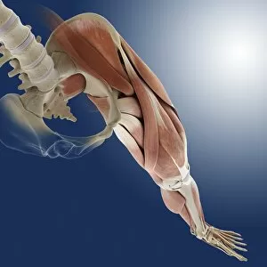
Leg muscles. Computer artwork of the bones and outer muscles of the left leg, seen from the front (anterior view) at an oblique angle looking downwards

Background imageMusculoskeletal System Collection: Thigh hamstring muscles, artwork C013 / 4428

Thigh hamstring muscles, artwork C013 / 4428
Thigh hamstring muscles. Computer artwork of a posterior view of two of the three hamstring muscles (red) of the thighs. The muscles are the semimembranosus (inner) and semitendinosus (outer)

Background imageMusculoskeletal System Collection: Iliopsoas muscles, artwork C013 / 4425

Iliopsoas muscles, artwork C013 / 4425
Iliopsoas muscles. Computer artwork of an anterior (frontal) view of the hips, showing the inner hip muscles (red), the ilioposas

Background imageMusculoskeletal System Collection: Buttock muscles, artwork C013 / 4423

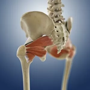
Buttock muscles, artwork C013 / 4423
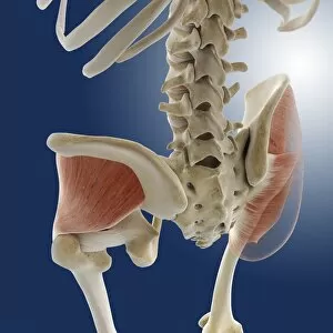
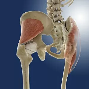
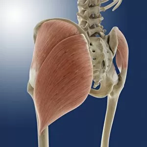
Buttock muscles. Computer artwork of a posterior oblique view of the hips, showing some of the muscles (red) of the buttocks

Background imageMusculoskeletal System Collection: Buttock muscles, artwork C013 / 4424

Buttock muscles, artwork C013 / 4424
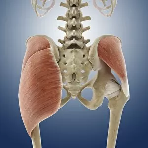
Buttock muscles. Computer artwork of a posterior view of the hips showing some of the muscles (red) of the buttocks. These are shown attached to the femurs (thigh bones) and the pelvis bones

Background imageMusculoskeletal System Collection: Buttock muscles, artwork C013 / 4422

Buttock muscles, artwork C013 / 4422
Buttock muscles. Computer artwork of a posterior oblique view of the hips showing some of the muscles (red) of the buttocks. These are shown attached to the femurs (thigh bones) and the pelvis bones

Background imageMusculoskeletal System Collection: Buttock muscles, artwork C013 / 4420

Buttock muscles, artwork C013 / 4420
Buttock muscles. Computer artwork of a posterior view of the hips showing some of the muscles (red) of the buttocks. These are shown attached to the femurs (thigh bones) and the pelvis bones

Background imageMusculoskeletal System Collection: Buttock muscles, artwork C013 / 4421

Buttock muscles, artwork C013 / 4421
Buttock muscles. Computer artwork of a side view (front of body at left) of the hips showing some of the muscles (red) of the buttocks

Background imageMusculoskeletal System Collection: Buttock muscles, artwork C013 / 4419

Buttock muscles, artwork C013 / 4419
Buttock muscles. Computer artwork of a posterior oblique view of the hips showing some of the muscles (red) of the buttocks. These are shown attached to the femurs (thigh bones) and the pelvis bones

Background imageMusculoskeletal System Collection: Buttock muscles, artwork C013 / 4415

Buttock muscles, artwork C013 / 4415
Buttock muscles. Computer artwork of a posterior oblique view of the hips showing some of the muscles (red) of the buttocks. These are shown attached to the femurs (thigh bones) and the pelvis bones

Background imageMusculoskeletal System Collection: Buttock muscles, artwork C013 / 4416

Buttock muscles, artwork C013 / 4416
Buttock muscles. Computer artwork of a posterior oblique view of the hips showing some of the muscles (red) of the buttocks. These are shown attached to the femurs (thigh bones) and the pelvis bones

Background imageMusculoskeletal System Collection: Osteopathy

Osteopathy. Osteopath manipulating a patients hand, using a thrust articular technique. Osteopathy uses massage and manipulation to treat a range of disorders

Background imageMusculoskeletal System Collection: Hip joint examination

Hip joint examination
MODEL RELEASED. Hip joint examination. Physiotherapist performing an assessment of the hip joints of a young boy. The bones of the hip are held together by ligaments

Background imageMusculoskeletal System Collection: Cervical spine examination

Cervical spine examination
MODEL RELEASED. Cervical spine examination. Physiotherapist manipulating the cervical (neck) region of a young boys spine, during a spinal examination

Background imageMusculoskeletal System Collection: Foot and ankle examination

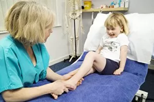
Foot and ankle examination
MODEL RELEASED. Foot and ankle examination. Physiotherapist preparing to perform a foot and ankle examination on a young child

Background imageMusculoskeletal System Collection: Upper spine examination

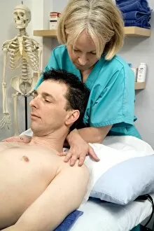
Upper spine examination
MODEL RELEASED. Upper spine examination. Physiotherapist manipulating the thoracic (upper) region of a patients spine, during a spinal examination

Background imageMusculoskeletal System Collection: Shoulder examination

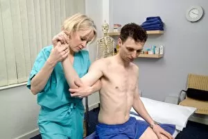
Shoulder examination
MODEL RELEASED. Shoulder examination. Physiotherapist manipulating a patients arm during an examination of his shoulder joint. This is known as a glenohumeral examination

Background imageMusculoskeletal System Collection: Stretching of trapezius back muscles

Stretching of trapezius back muscles
MODEL RELEASED. Stretching of trapezius back muscles. Physiotherapist using her crossed arms to raise a patients head and stretch the trapezius muscles of his upper back

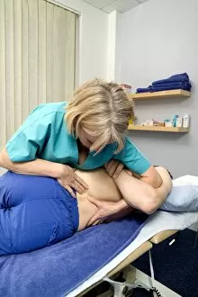
Background imageMusculoskeletal System Collection: Lumbar spine mobilisation treatment

Lumbar spine mobilisation treatment
MODEL RELEASED. Lumbar spine mobilisation treatment. Physiotherapist manipulating the lumbar (lower) region of a patients spine, during a spinal mobilisation treatment session

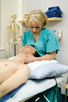
Background imageMusculoskeletal System Collection: Cervical spine mobilisation treatment

Cervical spine mobilisation treatment
MODEL RELEASED. Cervical spine mobilisation treatment. Physiotherapist manipulating the cervical (neck) region of a patients spine, during a spinal mobilisation treatment session

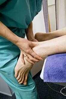
Background imageMusculoskeletal System Collection: Achillies tendon examination

Achillies tendon examination
MODEL RELEASED. Achillies tendon examination. Physiotherapist using her hands to examine and treat a patients Achilles tendon

Background imageMusculoskeletal System Collection: Apleys knee test

Apleys knee test
MODEL RELEASED. Apleys knee test. Physiotherapist performing an Apleys knee test to assess a patients knee injury. This test is carried out to diagnose knee cartilage injuries

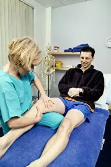
Background imageMusculoskeletal System Collection: Lachman knee ligament test

Lachman knee ligament test
MODEL RELEASED. Lachman knee ligament test. Physiotherapist performing a Lachman test on a patients knee ligaments. This test is designed as an assessment of the knees anterior cruciate ligament

Background imageMusculoskeletal System Collection: Ultrasound treatment of ligament injury

Ultrasound treatment of ligament injury
MODEL RELEASED. Ultrasound treatment of ligament injury. Ultrasound transducer (centre right) being used by a physiotherapist to treat a patients knee ligament injury

Background imageMusculoskeletal System Collection: McMurrays knee joint test

McMurrays knee joint test
MODEL RELEASED. McMurrays knee joint test. Physiotherapist performing a McMurrays knee joint test on a patient. The McMurray test is designed to assess injuries to the cartilage in the knee


For sale as Licensed Images

Choose your image, Select your licence and Download the media

The musculoskeletal system is a complex network of bones, muscles, ligaments, and nerves that work together to provide support and movement for the body. From the outer ankle ligaments to the inner ankle ligaments, every part plays a crucial role in maintaining stability and flexibility. Artwork C013 / 4452 beautifully depicts the intricate structure of the outer ankle ligaments, while artwork C013 / 4451 showcases the delicate nature of the inner ankle ligaments. These illustrations serve as a visual reminder of how these ligaments enable us to walk, run, and perform various activities with ease. Moving up towards the female muscular system artwork, we witness the strength and power that lies within our muscles. The human upper body illustration further emphasizes this by showcasing not only bones but also muscles and even our circulatory system. It reminds us that without proper muscle function, our bodies would lack mobility and vitality. Delving deeper into specific areas of our body's anatomy, we come across artwork depicting neck muscles and nerves (artwork), upper arm muscles (artwork), phrenic nerves alongside diaphragm (artwork), suboccipital muscles along with nerve (artwork C014 / 5097). These illustrations highlight how each muscle group works in harmony with its corresponding nerve supply to facilitate movement or maintain posture. Artworks such as arm anatomy (C013 / 4583) showcase detailed structures like tendons connecting muscle to bone while buttock muscles (C013 / 4414) remind us that even seemingly insignificant areas play vital roles in everyday movements like sitting or standing. Lastly, a cross-section biomedical illustration provides an overall view of an adult male's musculoskeletal system. This comprehensive representation serves as a reminder that all these individual parts are interconnected - from bones providing structural support to muscles enabling motion - creating an intricate web necessary for optimal functioning.