Nerve Cell Collection (#8)

The intricate world of nerve cells, also known as neurons, is a fascinating realm within our bodies

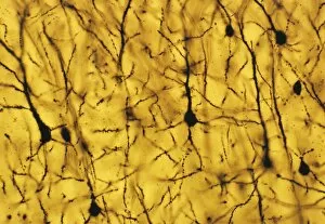
Background imageNerve Cell Collection: Foetal neurons

Foetal neurons. Light micrograph of human foetal neurons (nerve cells). Neurons are responsible for passing information around the central nervous system (CNS)

Background imageNerve Cell Collection: Nerve support cell, SEM

Nerve support cell, SEM
Nerve support cell. Coloured scanning electron micrograph (SEM) of an oligodendrocyte cell. This cell forms the myelin sheaths around nerve fibres in the central nervous system (brain and spinal cord)

Background imageNerve Cell Collection: Nerve cell growth

Nerve cell growth. Fluorescent light micrograph of a PC12 cell following stimulation by nerve growth factor. The cell body contains the nucleus (green)

Background imageNerve Cell Collection: Cultured nerve cells

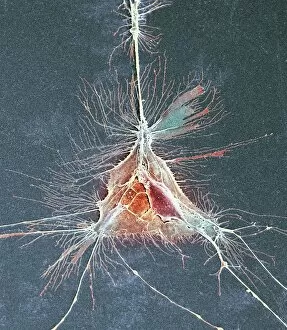
Cultured nerve cells. Coloured scanning electron micrograph (SEM) of a cultured piece of spinal cord (centre). Each nerve cell (neuron) in the cord has an axon (long thin strand) growing from it

Background imageNerve Cell Collection: Nerve cell culture, SEM

Nerve cell culture, SEM
Nerve cell culture. Coloured scanning electron micrograph (SEM) of new growth from a cultured sample from a spinal cord. The numerous branching strands are neurites

Background imageNerve Cell Collection: Cerebral cortex nerve cells

Cerebral cortex nerve cells

Background imageNerve Cell Collection: Purkinje nerve cell, SEM

Purkinje nerve cell, SEM

Background imageNerve Cell Collection: Granule nerve cell, SEM

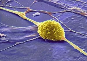
Granule nerve cell, SEM
Granule nerve cell. Coloured scanning electron micrograph (SEM) of a granule nerve cell (yellow) from the cerebellum of the brain

Background imageNerve Cell Collection: Purkinje nerve cell

Purkinje nerve cell

Background imageNerve Cell Collection: Purkinje nerve cells, SEM

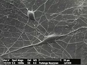
Purkinje nerve cells, SEM
Purkinje nerve cells. Scanning electron micrograph (SEM) of two Purkinje nerve cells from the cerebellum of the brain. The cells comprise a flask-shaped cell body

Background imageNerve Cell Collection: Unmyelinated intestinal nerve fibre, TEM

Unmyelinated intestinal nerve fibre, TEM
Unmyelinated intestinal nerve fibre. Coloured transmission electron micrograph (TEM) of a section across an nerve fibre from the intestine

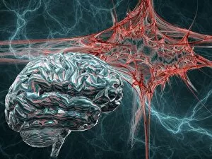
Background imageNerve Cell Collection: Brain and lightning, artwork

Brain and lightning, artwork
Brain and lightning. Computer artwork of a brain, a nerve cell (red) and lightning. This could represent electrical activity, thoughts and ideas, or an epileptic attack

Background imageNerve Cell Collection: Brain power

Brain power. Conceptual computer artwork of a Rubiks cube with neurological images on its surfaces. At top is a coloured magnetic resonance imaging (MRI) scan of a section through the head

Background imageNerve Cell Collection: Artwork showing action of sleeping drugs

Artwork showing action of sleeping drugs
Sleeping drug action. Artwork showing the action of various sleeping drugs on a nerve cell. The cell membrane (pink) is shown, along with associated proteins (orange)

Background imageNerve Cell Collection: Pain, conceptual computer artwork

Pain, conceptual computer artwork
Pain. Conceptual computer artwork of pain, where the site of pain is represented by a ball of barbed wire. Connected to this are the free nerve endings, which respond to mechanical

Background imageNerve Cell Collection: Artwork depicting Parkinsons disease

Artwork depicting Parkinsons disease

Background imageNerve Cell Collection: Demyelinated nerve, TEM

Demyelinated nerve, TEM
Demyelinated nerve. Coloured transmission electron micrograph (TEM) of a section through an axon (a structure that transmits nerve impulses to other nerve cells) that has lost its myelin sheath

Background imageNerve Cell Collection: Demyelinated nerve in multiple sclerosis

Demyelinated nerve in multiple sclerosis. Coloured transmission electron micrograph (TEM) of a section through an axon (a structure that transmits nerve impulses) that has lost its myelin sheath

Background imageNerve Cell Collection: Alzheimers disease brain cell, TEM

Alzheimers disease brain cell, TEM
Alzheimers disease brain cell. Coloured transmission electron micrograph (TEM) of a neurofibrillary tangle in a nerve cell from the brain of a patient with Alzheimers disease

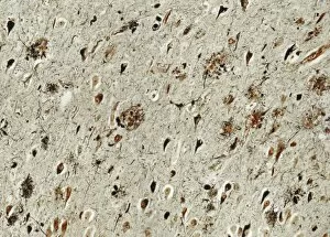
Background imageNerve Cell Collection: Alzheimers disease brain tissue

Alzheimers disease brain tissue, light micrograph. Two characteristic features of Alzheimers disease are seen here; neurofibrillary tangles (dark teardrop shapes)

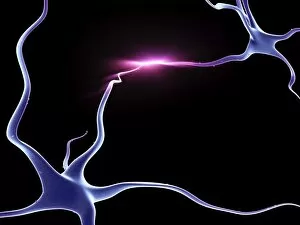
Background imageNerve Cell Collection: Nerve cells, neurons connected

Nerve cells, neurons connected
Computer artwork of two nerve cells connecting with glowing impulse

Background imageNerve Cell Collection: Ginkgo and nerve cells

Ginkgo and nerve cells
Ginkgo leaf and nerve cells. Computer artwork of a ginkgo (Ginkgo biloba) leaf with nerve cells. Ginkgo extracts are used in complementary medicine to aid concentration

Background imageNerve Cell Collection: Brain, neural network

Brain, neural network
Neural network. Computer artwork of a brain in top view, with the brains neural network represented by lines and flashes. A neural network is made up of nerve cells (neurons)

Background imageNerve Cell Collection: Alzheimers disease

Alzheimers disease
Conceptual computer artwork depicting Alzheimers disease and other brain disease


For sale as Licensed Images

Choose your image, Select your licence and Download the media

The intricate world of nerve cells, also known as neurons, is a fascinating realm within our bodies. These specialized cells play a crucial role in transmitting information throughout the nervous system. In the cerebellum tissue, under the lens of a light micrograph, we can observe the complex network formed by nerve and glial cells. This interplay between different cell types ensures proper functioning and communication within this region of the brain. Zooming in further with a transmission electron microscope (TEM), we witness the mesmerizing synapse nerve junctions - where two nerve cells meet to exchange vital signals. The intricacy of these connections highlights their importance in relaying messages across our neural pathways. Switching gears to scanning electron microscopy (SEM), we get an up-close look at individual nerve cells themselves. Their elongated structures and branching extensions showcase their ability to transmit electrical impulses efficiently. Moving on to hippocampus brain tissue, another essential area for memory formation and learning, we encounter Purkinje nerve cells nestled within the cerebellum. These large neurons have distinctive dendritic trees that receive inputs from various sources, contributing to motor coordination. As we explore further into brain tissue's complexity, it becomes evident that blood supply plays a crucial role in nourishing these delicate neural networks. A healthy flow ensures optimal functioning of all interconnected regions. Venturing beyond natural tissues into neural stem cell culture reveals exciting possibilities for regenerative medicine and understanding neurodevelopmental processes better. These cultured stem cells hold immense potential for repairing damaged nerves or studying neuronal growth patterns. Finally, let us not forget about cerebral cortex nerve cells - responsible for higher cognitive functions such as perception and decision-making. Their intricate arrangements enable us to process information effectively while navigating through daily life challenges. Whether observing cerebellum tissue or exploring neural stem cell cultures or marveling at synaptic connections under various microscopes – each glimpse into the world of nerve cells unveils new layers of complexity and highlights their indispensable role in our intricate neural symphony.