Nerve Cell Collection (#5)

The intricate world of nerve cells, also known as neurons, is a fascinating realm within our bodies

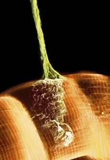
Background imageNerve Cell Collection: Synapse, SEM

Synapse, SEM
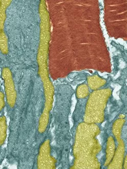
Synapse. Coloured scanning electron micrograph (SEM) of a neuromuscular junction showing a motor neurone (green) terminating on skeletal muscle fibres (orange)

Background imageNerve Cell Collection: Induced nerve stem cells, micrograph

Induced nerve stem cells, micrograph
Induced nerve stem cells. Fluorescence light micrograph of neural (nerve) stem cells that have been created (induced) from human adult skin fibroblast cells by gene manipulation

Background imageNerve Cell Collection: Stem cell-derived nerve cells

Stem cell-derived nerve cells. Fluorescence light micrograph of neural (nerve) stem cells that have been derived from human embryonic stem cells (HESC)

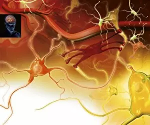
Background imageNerve Cell Collection: Brain cells, artwork

Brain cells, artwork
Brain cells. Computer artwork representing neurons (nerve cells) in the brain

Background imageNerve Cell Collection: Brain cell, TEM C014 / 0358

Brain cell, TEM C014 / 0358
Brain cell. Transmission electron micrograph (TEM) of a section through an oligodendrocyte in human brain tissue. Oligodendrocytes occur in both the white

Background imageNerve Cell Collection: Brain cell, TEM C014 / 0359

Brain cell, TEM C014 / 0359
Brain cell. Transmission electron micrograph (TEM) of a section through an oligodendrocyte in human brain tissue. Oligodendrocytes occur in both the white

Background imageNerve Cell Collection: Deep brain stimulation and nerve cell C016 / 7205

Deep brain stimulation and nerve cell C016 / 7205
Deep brain stimulation. Computer artwork showing an electrode sending electrical impulses to nerve cells. In the background a light micrograph (LM) of a section through grey matter in the brain

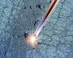
Background imageNerve Cell Collection: Deep brain stimulation and nerve cell C016 / 7203

Deep brain stimulation and nerve cell C016 / 7203
Deep brain stimulation. Computer artwork showing an electrode sending electrical impulses to nerve cells. In the background a light micrograph (LM) of a section through grey matter in the brain

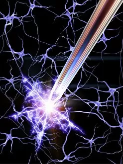
Background imageNerve Cell Collection: Deep brain stimulation and nerve cells C016 / 7206

Deep brain stimulation and nerve cells C016 / 7206
Deep brain stimulation (DBS). Computer artwork showing an electrode sending electrical impulses to nerve cells. DBS was developed for the treatment of Parkinsons disease

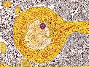
Background imageNerve Cell Collection: Purkinje nerve cell, TEM C014 / 0582

Purkinje nerve cell, TEM C014 / 0582
Purkinje nerve cell. Transmission electron micrograph (TEM) of a purkinje nerve cell (bright yellow, centre) from the cerebellum of the brain

Background imageNerve Cell Collection: Nerve cells, artwork C018 / 2887

Nerve cells, artwork C018 / 2887
Nerve cells, or neurons, computer artwork. Neurons are responsible for passing information around the central nervous system (CNS) and from the CNS to the rest of the body

Background imageNerve Cell Collection: Neural network, conceptual image C013 / 9958

Neural network, conceptual image C013 / 9958
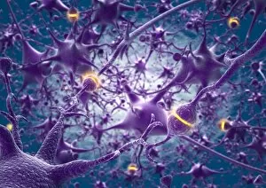
Neural network, conceptual image. Computer artwork representing interconnecting nerve cells (neurons)

Background imageNerve Cell Collection: Neural network, conceptual image C013 / 9957

Neural network, conceptual image C013 / 9957
Neural network, conceptual image. Computer artwork representing interconnecting nerve cells (neurons)

Background imageNerve Cell Collection: Nerve cell, SEM C013 / 9772

Nerve cell, SEM C013 / 9772
Nerve cell. Coloured scanning electron micrograph (SEM) of a nerve cell (neuron). Neurons are responsible for passing information around the central nervous system (CNS)

Background imageNerve Cell Collection: Cerebral cortex nerve cells C013 / 9767

Cerebral cortex nerve cells C013 / 9767
Cerebral cortex nerve cells. Light micrograph of a section through neurones (nerve cells, black) in the cerebral cortex of a human brain

Background imageNerve Cell Collection: Purkinje nerve cell C013 / 9763

Purkinje nerve cell C013 / 9763
Purkinje nerve cell. Light micrograph of a purkinje nerve cell (orange, centre) from the cerebellum of the brain. The cell comprises a flask-shaped cell body from which numerous processes (dendrites)

Background imageNerve Cell Collection: Purkinje nerve cells C013 / 9745

Purkinje nerve cells C013 / 9745
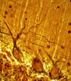
Purkinje nerve cells. Light micrograph of three purkinje nerve cells (across bottom) from the cerebellum of the brain. The cells comprise a flask-shaped cell body from which numerous processes

Background imageNerve Cell Collection: Neural network, artwork C013 / 4636

Neural network, artwork C013 / 4636
Neural network. Computer artwork of nerve cells (neurons) connected by processes (filaments), known as dendrites and axons, to form a neural network

Background imageNerve Cell Collection: Retinal rod cell, TEM C013 / 4805

Retinal rod cell, TEM C013 / 4805
Retinal rod cell. Transmission electron micrograph (TEM) of a section through a rod cell from the retina of an eye, showing the inner segment (bottom) filled with mitochondria (green)

Background imageNerve Cell Collection: Retinal rod cell, TEM C013 / 4804

Retinal rod cell, TEM C013 / 4804
Retinal rod cell. Transmission electron micrograph (TEM) of a section through a rod cell from the retina of an eye, showing the inner segment (bottom) filled with mitochondria (green)

Background imageNerve Cell Collection: Brain cells, TEM C013 / 4801

Brain cells, TEM C013 / 4801
Brain cells. Transmission electron micrograph (TEM) of a section through oligodendrocytes (dark) in human brain tissue, showing free ribosomes (dark green dots), golgi apparatus (curved brown lines)

Background imageNerve Cell Collection: Macrophage engulfing a nerve cell, TEM C013 / 4803

Macrophage engulfing a nerve cell, TEM C013 / 4803
Macrophage engulfing a nerve cell. Transmission electron micrograph (TEM) of a section through a macrophage white blood cell (blue) that has engulfed a nerve cell (neuron, centre)

Background imageNerve Cell Collection: Brain cells, TEM C013 / 4800

Brain cells, TEM C013 / 4800
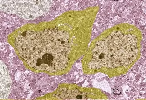
Brain cells. Transmission electron micrograph (TEM) of a section through oligodendrocytes in human brain tissue, showing free ribosomes (dark pink dots), golgi apparatus (curved brown lines)

Background imageNerve Cell Collection: Nerve cell, TEM C013 / 4797

Nerve cell, TEM C013 / 4797
Nerve cell. Transmission electron micrograph (TEM) of a section through a neuron (nerve cell), showing characteristic Nissl body (dark blue lines), numerous golgi apparatus (curved green lines)

Background imageNerve Cell Collection: Brain cell, TEM C013 / 4799

Brain cell, TEM C013 / 4799
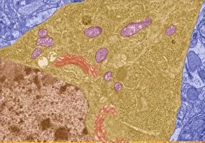
Brain cell. Transmission electron micrograph (TEM) of a section through an oligodendrocyte in human brain tissue, showing free ribosomes (dark brown dots), golgi apparatus (curved orange lines)

Background imageNerve Cell Collection: Nerve cell, TEM C013 / 4796

Nerve cell, TEM C013 / 4796
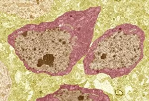
Nerve cell. Transmission electron micrograph (TEM) of a section through a neuron (nerve cell), showing characteristic Nissl body (dark red lines), numerous golgi apparatus (curved pink lines)

Background imageNerve Cell Collection: Brain cell, TEM C013 / 4798

Brain cell, TEM C013 / 4798
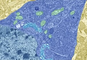
Brain cell. Transmission electron micrograph (TEM) of a section through an oligodendrocyte in human brain tissue, showing free ribosomes (dark blue dots), golgi apparatus (curved light blue lines)

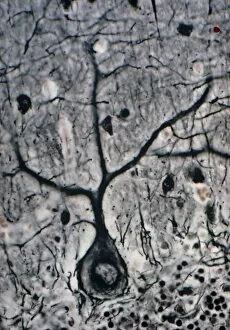
Background imageNerve Cell Collection: LM of a Purkinje cell in the cerebellum

LM of a Purkinje cell in the cerebellum

Background imageNerve Cell Collection: False-colour SEM of 3 neurones of cerebral cortex

False-colour SEM of 3 neurones of cerebral cortex
False-colour scanning electron micrograph (SEM) showing three neurones (nerve cells) of the human cerebral cortex - the outer grey matter of the brain

Background imageNerve Cell Collection: Brain nerve cells

Brain nerve cells
Nerve cells in the brain. Artwork showing the different types of nerve cells in the grey matter of the brain. Neurons (yellow, for example at lower right) relay nerve signals around the brain

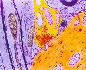
Background imageNerve Cell Collection: Myelin surrounding a nerve axon, TEM

Myelin surrounding a nerve axon, TEM
Myelin surrounding a nerve axon, coloured transmission electron micrograph (TEM). The concentric round rings are the sheets of a Schwann cells myelin membrane (brown rings)

Background imageNerve Cell Collection: Artwork of a nerve cell of the brain & a synapse

Artwork of a nerve cell of the brain & a synapse
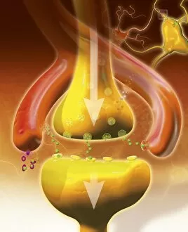
Nerve cell and synapses. Artwork of a nerve cell (neurone) of the brain with a close-up of a synapse. The nerve cell body is at lower right

Background imageNerve Cell Collection: Motor neurone nerve cell and synapses

Motor neurone nerve cell and synapses
Motor neurone and synapses. Computer graphic of a motor neurone and synapses (glowing yellow bulges, at lower centre). The cell body is at centre

Background imageNerve Cell Collection: False-col SEM of single neurone of cerebral cortex

False-col SEM of single neurone of cerebral cortex
False-colour scanning electron micrograph (SEM) of a single neurone (nerve cell) of the human cerebral cortex - the outer grey matter of the brain

Background imageNerve Cell Collection: Oligodendrocyte and microglia brain cells

Oligodendrocyte and microglia brain cells
Oligodendrocyte and microglia action in the brain. At top, the axon (output process, orange) of a neuron (nerve cell) is seen

Background imageNerve Cell Collection: Computer graphic of a motor neuron nerve cell

Computer graphic of a motor neuron nerve cell
Motor neurone. Computer graphic of a motor neuron nerve cell. At centre is the cell body; at the centre of this is a red circle which is the cell nucleus

Background imageNerve Cell Collection: LM of cortex and medulla of the cerebellum

LM of cortex and medulla of the cerebellum
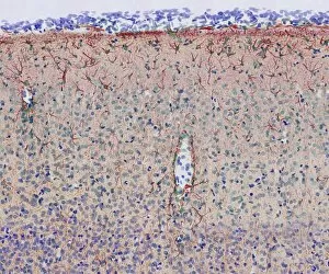
Cerebellum. Light micrograph of a cross-section through the cortex (surface) and medulla (core) of a human cerebellum, part of the brain

Background imageNerve Cell Collection: Artwork showing numerous multipolar neurones

Artwork showing numerous multipolar neurones
Illustration based on a scanning electron micrograph (SEM) of numerous multipolar neurones (nerve cells) growing in tissue culture

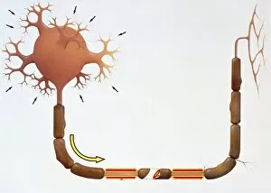
Background imageNerve Cell Collection: Artwork of human motor neuron (nerve cell)

Artwork of human motor neuron (nerve cell)
Motor neuron. Illustration of a human motor neuron (nerve cell). The arrows show the direction of the nervous impulse, which enters the cell body (top left) through minute processes called dendrites

Background imageNerve Cell Collection: Coloured TEM of a nerve synapse

Coloured TEM of a nerve synapse

Background imageNerve Cell Collection: LM of purkinje nerve cells in the cerebellum

LM of purkinje nerve cells in the cerebellum
Purkinje nerve cells. Light micrograph of a row of Purkinje nerve cells from the cerebellum of the human brain. Each Purkinje cell is composed of a flask-shaped cell body

Background imageNerve Cell Collection: LM of nerve cells and fibres in brain tissue

LM of nerve cells and fibres in brain tissue
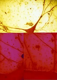
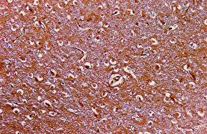
Brain tissue. Light micrograph of a section through the grey matter of a normal brain. There are several nerve cell bodies (objects in white areas)

Background imageNerve Cell Collection: Light micrograph of a nerve cell

Light micrograph of a nerve cell
Neuron. Scanning electron micrograph of a nerve cell with a branching axon. On the right is the cell body, which contains the nucleus

Background imageNerve Cell Collection: Purkinje nerve cells

Purkinje nerve cells

Background imageNerve Cell Collection: Coloured computer image of a nerve cell

Coloured computer image of a nerve cell
Nerve cell. Coloured computer image based on laser scanning microscope sections through a nerve cell. Many dendrites (branching projections) can be seen extending from the bulbous cell body

Background imageNerve Cell Collection: Brain cortex tissue, light micrograph

Brain cortex tissue, light micrograph
Brain cortex tissue. Light micrograph of a section through tissue from the cortex of the brain. The cortex, or grey matter, is the outer layer of the brain

Background imageNerve Cell Collection: Astrocyte / neuron synapse in the brain

Astrocyte / neuron synapse in the brain
Astrocyte/neuron synapse in the brain. Artwork of a nerve cell (neuron, yellow) synapse in the brain with an associated regulatory astrocyte cell (red)

Background imageNerve Cell Collection: Retina blood vessel and nerve cells

Retina blood vessel and nerve cells
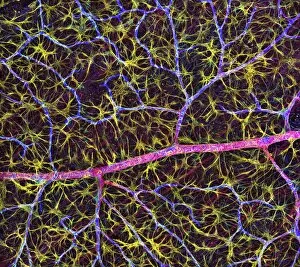
Retina cells. Fluorescent light micrograph of cells in the retina, the light-sensitive membrane that lines the back of the eyeball


For sale as Licensed Images

Choose your image, Select your licence and Download the media

The intricate world of nerve cells, also known as neurons, is a fascinating realm within our bodies. These specialized cells play a crucial role in transmitting information throughout the nervous system. In the cerebellum tissue, under the lens of a light micrograph, we can observe the complex network formed by nerve and glial cells. This interplay between different cell types ensures proper functioning and communication within this region of the brain. Zooming in further with a transmission electron microscope (TEM), we witness the mesmerizing synapse nerve junctions - where two nerve cells meet to exchange vital signals. The intricacy of these connections highlights their importance in relaying messages across our neural pathways. Switching gears to scanning electron microscopy (SEM), we get an up-close look at individual nerve cells themselves. Their elongated structures and branching extensions showcase their ability to transmit electrical impulses efficiently. Moving on to hippocampus brain tissue, another essential area for memory formation and learning, we encounter Purkinje nerve cells nestled within the cerebellum. These large neurons have distinctive dendritic trees that receive inputs from various sources, contributing to motor coordination. As we explore further into brain tissue's complexity, it becomes evident that blood supply plays a crucial role in nourishing these delicate neural networks. A healthy flow ensures optimal functioning of all interconnected regions. Venturing beyond natural tissues into neural stem cell culture reveals exciting possibilities for regenerative medicine and understanding neurodevelopmental processes better. These cultured stem cells hold immense potential for repairing damaged nerves or studying neuronal growth patterns. Finally, let us not forget about cerebral cortex nerve cells - responsible for higher cognitive functions such as perception and decision-making. Their intricate arrangements enable us to process information effectively while navigating through daily life challenges. Whether observing cerebellum tissue or exploring neural stem cell cultures or marveling at synaptic connections under various microscopes – each glimpse into the world of nerve cells unveils new layers of complexity and highlights their indispensable role in our intricate neural symphony.