Neurological Collection (#4)

Neurological wonders unravel before our eyes as we delve into the intricate world of the brain

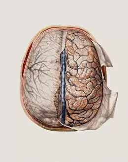
Background imageNeurological Collection: Brain meninges

Brain meninges, historical anatomical artwork. This cranial (top) view of the brain (with front at the top) and its surrounding protective meninges, which have been partially dissected away

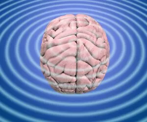
Background imageNeurological Collection: Brain waves, conceptual image

Brain waves, conceptual image
Brain waves. Conceptual image of waves emanating from a human brain. This can represent the electrical activity of the brain, as recorded on an electroencephalogram (EEG)

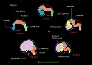
Background imageNeurological Collection: Foetal brain development, artwork

Foetal brain development, artwork. During the 4th week (upper left) the neural tube begins to differentiate into a spinal cord (green), forebrain (blue), midbrain (grey) and hindbrain (orange)

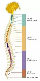
Background imageNeurological Collection: Spinal cord regions, diagram

Spinal cord regions, diagram
Spinal cord regions. Diagram of a sagittal section through the human skull and backbone, showing the five main regions of the spinal cord and the numbered nerves associated with them

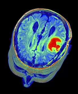
Background imageNeurological Collection: Multiple sclerosis, MRI scan

Multiple sclerosis, MRI scan

Background imageNeurological Collection: Brain and spinal cord, artwork

Brain and spinal cord, artwork
Brain and spinal cord. Computer artwork showing the brain (top) and segments of the spinal cord. The spinal cord begins at the medulla oblongata at the base of the brainstem

Background imageNeurological Collection: Brain tumour, 3D-MRI scan

Brain tumour, 3D-MRI scan
Brain tumour. Coloured three-dimensional model of a male head, made up from numerous magnetic resonance imaging (MRI) scans

Background imageNeurological Collection: Charles Bell (1774-1842) Scottish surgeon and anatomist: neurological discoveries

Charles Bell (1774-1842) Scottish surgeon and anatomist: neurological discoveries: Bells Palsy (a facial paralysis) named after him. Wood engraving

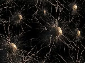
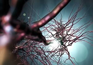
Background imageNeurological Collection: Astrocyte nerve cells, artwork C017 / 3425

Astrocyte nerve cells, artwork C017 / 3425
Astrocyte nerve cells, computer artwork. Astrocytes are a type of glial cell.They provide structural support and protection for neurons (nerve cells) and also supply them with nutrients and oxygen

Background imageNeurological Collection: Pyramidal nerve cells, artwork C017 / 2273

Pyramidal nerve cells, artwork C017 / 2273
Pyramidal nerve cells. Computer artwork of pyramidal nerve cells from the cerebral cortex of the brain. Pyramidal cells are so named for their triangular cell bodies

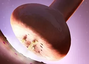
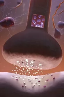
Background imageNeurological Collection: Nerve synapse, artwork C017 / 3427

Nerve synapse, artwork C017 / 3427
Nerve synapse. Computer artwork of of a junction, or synapse, between two nerve cells (neurons). As the electrical signal reaches the presynaptic end of a neuron it triggers the release of

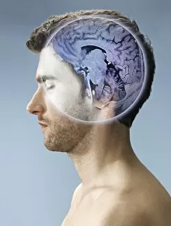
Background imageNeurological Collection: Brain scan, conceptual image C017 / 7392

Brain scan, conceptual image C017 / 7392
MODEL RELEASED. Brain scan, conceptual composite image

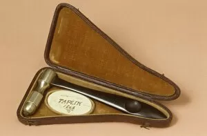
Background imageNeurological Collection: Bennet percussor, circa 1860 C017 / 6934

Bennet percussor, circa 1860 C017 / 6934
Bennet percussor with ivory pleximeter dating from 1860s, England. A percussor is a small hammer used for testing reflexes and for percussive examinations of the chest

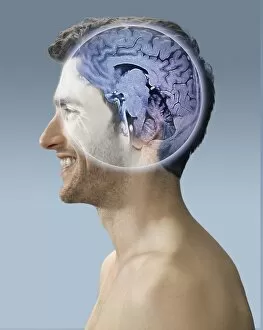
Background imageNeurological Collection: Brain scan, conceptual image C017 / 7394

Brain scan, conceptual image C017 / 7394
MODEL RELEASED. Brain scan, conceptual composite image

Background imageNeurological Collection: Brain scan, conceptual image C017 / 7398

Brain scan, conceptual image C017 / 7398
MODEL RELEASED. Brain scan, conceptual composite image

Background imageNeurological Collection: Brain scan, conceptual image C017 / 7397

Brain scan, conceptual image C017 / 7397
MODEL RELEASED. Brain scan, conceptual composite image

Background imageNeurological Collection: Bennet percussor, circa 1860 C017 / 6971

Bennet percussor, circa 1860 C017 / 6971
Bennet percussor dating from 1860s, England. A percussor is a small hammer used for testing reflexes and for percussive examinations of the chest

Background imageNeurological Collection: Nerve synapse, artwork C017 / 3428

Nerve synapse, artwork C017 / 3428
Nerve synapse. Computer artwork of of a junction, or synapse, between two nerve cells (neurons). As the electrical signal reaches the presynaptic end of a neuron it triggers the release of

Background imageNeurological Collection: Corpus callosum, DTI MRI scan C017 / 7044

Corpus callosum, DTI MRI scan C017 / 7044
Corpus callosum. Coronal 3D diffusion tensor imaging (DTI) magnetic resonance imaging (MRI) scan of nerve pathways (coloured) in and around the brains corpus callosum

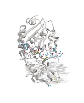
Background imageNeurological Collection: SIRT3 molecule, artwork C017 / 3657

SIRT3 molecule, artwork C017 / 3657
SIRT3 molecule. Computer artwork showing the structure of a molecule of NAD-dependent deacetylase sirtuin-3, mitochondrial (SIRT3)

Background imageNeurological Collection: Pyramidal nerve cells, artwork C017 / 2274

Pyramidal nerve cells, artwork C017 / 2274
Pyramidal nerve cells. Computer artwork of pyramidal nerve cells from the cerebral cortex of the brain. Pyramidal cells are so named for their triangular cell bodies

Background imageNeurological Collection: Brain scan, conceptual image C017 / 7391

Brain scan, conceptual image C017 / 7391
MODEL RELEASED. Brain scan, conceptual composite image

Background imageNeurological Collection: Synaesthesia, conceptual artwork C017 / 7072

Synaesthesia, conceptual artwork C017 / 7072
Synaesthesia, conceptual artwork. Synaesthesia is a neurological condition where stimulation of one sensory or cognitive pathway leads to automatic

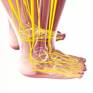
Background imageNeurological Collection: Human foot nervous system, artwork F007 / 1148

Human foot nervous system, artwork F007 / 1148
Human foot nervous system, computer artwork

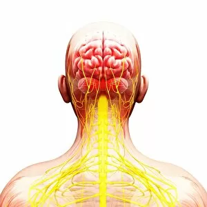
Background imageNeurological Collection: Female nervous system, artwork F007 / 4284

Female nervous system, artwork F007 / 4284
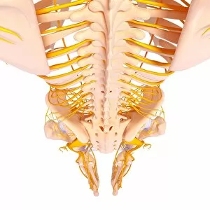
Female nervous system, computer artwork

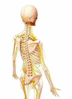
Background imageNeurological Collection: Female nervous system, artwork F007 / 3461

Female nervous system, artwork F007 / 3461
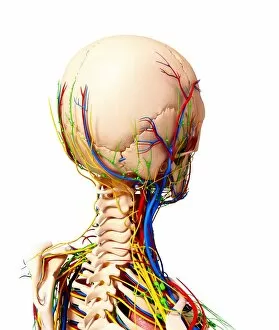
Female nervous system, computer artwork

Background imageNeurological Collection: Human nervous system, artwork F007 / 3986

Human nervous system, artwork F007 / 3986
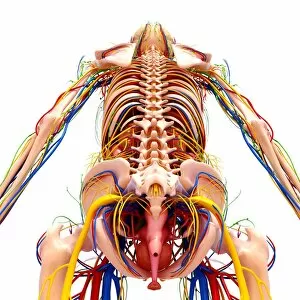
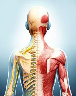
Human nervous system, computer artwork

Background imageNeurological Collection: Human nervous system, artwork F007 / 3394

Human nervous system, artwork F007 / 3394
Human nervous system, computer artwork

Background imageNeurological Collection: Female anatomy, artwork F007 / 3969

Female anatomy, artwork F007 / 3969
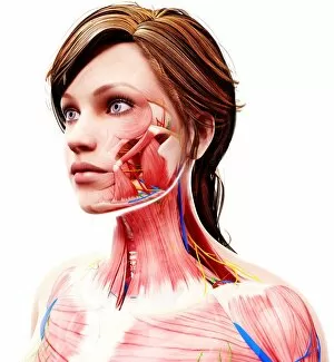
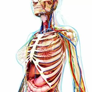
Female anatomy, computer artwork

Background imageNeurological Collection: Female anatomy, artwork F007 / 3665

Female anatomy, artwork F007 / 3665
Female anatomy, computer artwork

Background imageNeurological Collection: Human nervous system, artwork F007 / 4283

Human nervous system, artwork F007 / 4283
Human nervous system, computer artwork

Background imageNeurological Collection: Female nervous system, artwork F007 / 4298

Female nervous system, artwork F007 / 4298
Female nervous system, computer artwork

Background imageNeurological Collection: Human nervous system, artwork F007 / 4193

Human nervous system, artwork F007 / 4193
Human nervous system, computer artwork

Background imageNeurological Collection: Female anatomy, artwork F007 / 4084

Female anatomy, artwork F007 / 4084
Female anatomy, computer artwork

Background imageNeurological Collection: Female anatomy, artwork F007 / 3976

Female anatomy, artwork F007 / 3976
Female anatomy, computer artwork

Background imageNeurological Collection: Female nervous system, artwork F007 / 3589

Female nervous system, artwork F007 / 3589
Female nervous system, computer artwork

Background imageNeurological Collection: Human nervous system, artwork F007 / 4085

Human nervous system, artwork F007 / 4085
Human nervous system, computer artwork

Background imageNeurological Collection: Human anatomy, artwork F007 / 3686

Human anatomy, artwork F007 / 3686
Human anatomy, computer artwork

Background imageNeurological Collection: Female anatomy, artwork F007 / 3768

Female anatomy, artwork F007 / 3768
Female anatomy, computer artwork

Background imageNeurological Collection: Female anatomy, artwork F007 / 3868

Female anatomy, artwork F007 / 3868
Female anatomy, computer artwork

Background imageNeurological Collection: Human anatomy, artwork F007 / 3471

Human anatomy, artwork F007 / 3471
Human anatomy, computer artwork

Background imageNeurological Collection: Female nervous system, artwork F007 / 3870

Female nervous system, artwork F007 / 3870
Female nervous system, computer artwork

Background imageNeurological Collection: Female nervous system, artwork F007 / 3574

Female nervous system, artwork F007 / 3574
Female nervous system, computer artwork

Background imageNeurological Collection: Human anatomy, artwork F007 / 3463

Human anatomy, artwork F007 / 3463
Human anatomy, computer artwork

Background imageNeurological Collection: Female anatomy, artwork F007 / 3771

Female anatomy, artwork F007 / 3771
Female anatomy, computer artwork

Background imageNeurological Collection: Female anatomy, artwork F007 / 3565

Female anatomy, artwork F007 / 3565
Female anatomy, computer artwork

Background imageNeurological Collection: Human anatomy, artwork F007 / 4090

Human anatomy, artwork F007 / 4090
Human anatomy, computer artwork

Background imageNeurological Collection: Human nervous system, artwork F007 / 3378

Human nervous system, artwork F007 / 3378
Human nervous system, computer artwork


For sale as Licensed Images

Choose your image, Select your licence and Download the media

Neurological wonders unravel before our eyes as we delve into the intricate world of the brain. The Motor homunculus model, a representation of the body's motor functions within the brain, unveils the fascinating connection between mind and movement. Cerebellum tissue, captured in a light micrograph, showcases its remarkable structure responsible for coordination and balance. Through Brain fibres revealed by DTI MRI scan C017 / 7099, we witness the complex network that allows information to flow seamlessly throughout our minds. These Brain pathways pave the way for thoughts to travel and actions to be executed with precision. The Brain blood vessels depicted in a stunning 3D angiogram C007 / 1981 remind us of their vital role in nourishing this extraordinary organ. As we explore further, we encounter both Motor and sensory homunculi – visual representations highlighting how different regions of our bodies are represented within specific areas of our brains. Delving deeper into microscopic realms, Nerve and glial cells come alive under a light micrograph. Their intricate structures reveal their crucial roles in transmitting signals and supporting neuronal health. Synapse nerve junctions captured through TEM imagery demonstrate where these incredible cells communicate with one another. Surprisingly, even caffeine crystals make an appearance under a light microscope – reminding us that neurological effects can extend beyond what meets the eye. Zooming closer still reveals Nerve cells observed through SEM imaging; their unique shapes hint at their diverse functions within this awe-inspiring system. Finally, Hippocampus brain tissue takes center stage – known for its involvement in memory formation and spatial navigation. Its intricacies hold secrets waiting to be unlocked by dedicated researchers worldwide. As we uncover these neurological marvels piece by piece, it becomes evident that there is much more than meets the eye when it comes to understanding this enigmatic realm within each one of us - an endless source of fascination awaiting exploration.