Neurological Collection (#6)

Neurological wonders unravel before our eyes as we delve into the intricate world of the brain

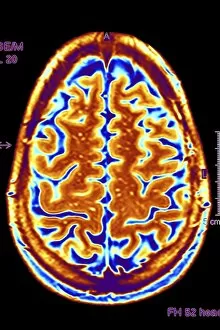
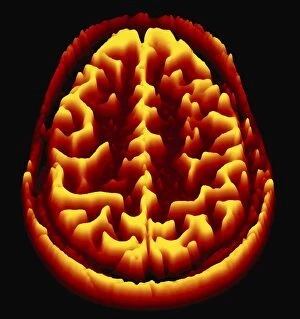
Background imageNeurological Collection: Brain scan, MRI scan, heightmap F006 / 7096

Brain scan, MRI scan, heightmap F006 / 7096
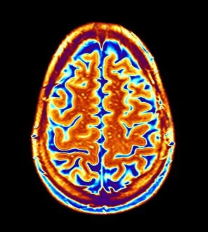
Normal brain. Coloured magnetic resonance imaging (MRI) scan of an axial section through a healthy brain, converted into a heightmap or height field

Background imageNeurological Collection: Brain activity, artwork F006 / 4599

Brain activity, artwork F006 / 4599
Brain activity. Computer artwork of EEG (electroencephalogram) traces superimposed over a brain illustration. An EEG records the brains activity

Background imageNeurological Collection: Brain scan, MRI scan, heightmap F006 / 7080

Brain scan, MRI scan, heightmap F006 / 7080
Normal brain. Coloured magnetic resonance imaging (MRI) scan of an axial section through a healthy brain, converted into a heightmap or height field

Background imageNeurological Collection: Cyclist, artwork F006 / 7385

Cyclist, artwork F006 / 7385
Cyclist, computer artwork

Background imageNeurological Collection: Brain scan, MRI scan F006 / 4629

Brain scan, MRI scan F006 / 4629
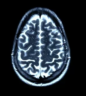
Normal brain. Coloured magnetic resonance imaging (MRI) scan of an axial section through a healthy brain. The front of the brain is towards top of the image

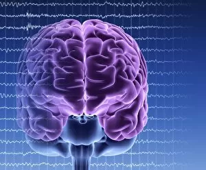
Background imageNeurological Collection: Brain activity, artwork F006 / 4626

Brain activity, artwork F006 / 4626
Brain activity. Computer artwork of EEG (electroencephalogram) traces superimposed over a brain illustration. An EEG records the brains activity

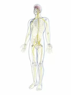
Background imageNeurological Collection: Male nervous system, artwork F006 / 7484

Male nervous system, artwork F006 / 7484
Male nervous system, computer artwork

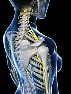
Background imageNeurological Collection: Female nervous system, artwork F006 / 7559

Female nervous system, artwork F006 / 7559
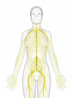
Female nervous system, computer artwork

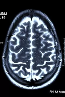
Background imageNeurological Collection: Brain scan, MRI scan F006 / 4610

Brain scan, MRI scan F006 / 4610
Normal brain. Coloured magnetic resonance imaging (MRI) scan of an axial section through a healthy brain. The front of the brain is towards top of the image

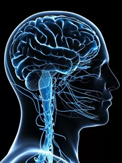
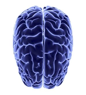
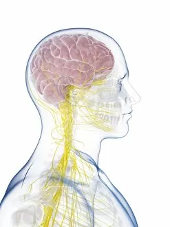
Background imageNeurological Collection: Male brain, artwork F006 / 7477

Male brain, artwork F006 / 7477
Male brain, computer artwork

Background imageNeurological Collection: Brain scan, MRI scan F006 / 4617

Brain scan, MRI scan F006 / 4617
Normal brain. Coloured magnetic resonance imaging (MRI) scan of an axial section through a healthy brain. The front of the brain is towards top of the image

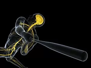
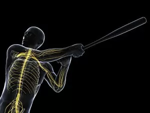
Background imageNeurological Collection: Baseball player, artwork F006 / 7315

Baseball player, artwork F006 / 7315
Baseball player, computer artwork

Background imageNeurological Collection: Male nervous system, artwork F006 / 7485

Male nervous system, artwork F006 / 7485
Male nervous system, computer artwork

Background imageNeurological Collection: MRI brain scan and nerve cells F006 / 3595

MRI brain scan and nerve cells F006 / 3595
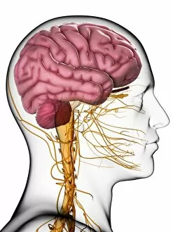
Computer artwork of a sagittal (side) view of a human head. The largest part of the brain is the cerebrum (upper part). This surrounds the limbic system (curved structures at upper centre)

Background imageNeurological Collection: Brain scan, MRI scan F006 / 4607

Brain scan, MRI scan F006 / 4607
Normal brain. Coloured magnetic resonance imaging (MRI) scan of an axial section through a healthy brain. The front of the brain is towards top of the image

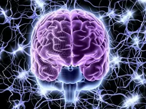
Background imageNeurological Collection: Brain and nerve cells, neural network F006 / 3585

Brain and nerve cells, neural network F006 / 3585
Computer artwork of a frontal view of a human brain. In the background a neural network of nerve cells firing

Background imageNeurological Collection: Baseball player, artwork F006 / 7310

Baseball player, artwork F006 / 7310
Baseball player, computer artwork

Background imageNeurological Collection: Human brain, artwork F006 / 6313

Human brain, artwork F006 / 6313
Human brain, computer artwork. The front of the brain is at bottom

Background imageNeurological Collection: Brain activity, artwork F006 / 4622

Brain activity, artwork F006 / 4622
Brain activity. Computer artwork of EEG (electroencephalogram) traces and a brain illustration. An EEG records the brains activity
Background imageNeurological Collection: Cyclist, artwork F006 / 7388

Cyclist, artwork F006 / 7388
Cyclist, computer artwork

Background imageNeurological Collection: Brain scan, MRI scan, heightmap F006 / 7078

Brain scan, MRI scan, heightmap F006 / 7078
Normal brain. Coloured magnetic resonance imaging (MRI) scan of an axial section through a healthy brain, converted into a heightmap or height field

Background imageNeurological Collection: Brain scan, MRI scan, heightmap F006 / 7093

Brain scan, MRI scan, heightmap F006 / 7093
Normal brain. Coloured magnetic resonance imaging (MRI) scan of an axial section through a healthy brain, converted into a heightmap or height field

Background imageNeurological Collection: Male nervous system, artwork F006 / 7486

Male nervous system, artwork F006 / 7486
Male nervous system, computer artwork

Background imageNeurological Collection: Brain activity, artwork F006 / 4604

Brain activity, artwork F006 / 4604
Brain activity. Computer artwork of EEG (electroencephalogram) traces and a brain illustration. An EEG records the brains activity
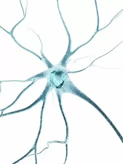
Background imageNeurological Collection: Male nervous system, artwork F006 / 7466

Male nervous system, artwork F006 / 7466
Male nervous system, computer artwork

Background imageNeurological Collection: Sleeping man, artwork F006 / 7362

Sleeping man, artwork F006 / 7362
Sleeping man, computer artwork

Background imageNeurological Collection: Brain activity, artwork F006 / 4630

Brain activity, artwork F006 / 4630
Brain activity. Computer artwork of EEG (electroencephalogram) traces superimposed over a brain illustration. An EEG records the brains activity
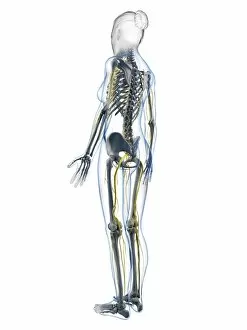
Background imageNeurological Collection: Brain scan, MRI scan, heightmap F006 / 7081

Brain scan, MRI scan, heightmap F006 / 7081
Normal brain. Coloured magnetic resonance imaging (MRI) scan of an axial section through a healthy brain, converted into a heightmap or height field

Background imageNeurological Collection: Sleeping man, artwork F006 / 7357

Sleeping man, artwork F006 / 7357
Sleeping man, computer artwork

Background imageNeurological Collection: Brain scan, MRI scan, heightmap F006 / 7091

Brain scan, MRI scan, heightmap F006 / 7091
Normal brain. Coloured magnetic resonance imaging (MRI) scan of an axial section through a healthy brain, converted into a heightmap or height field
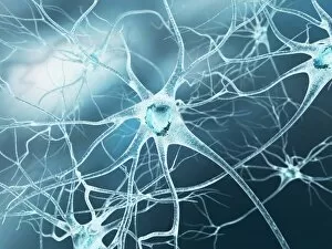
Background imageNeurological Collection: Female anatomy, artwork F007 / 6956

Female anatomy, artwork F007 / 6956
Female skeleton and nervous system, computer artwork

Background imageNeurological Collection: Male anatomy, artwork F007 / 7364

Male anatomy, artwork F007 / 7364
Male anatomy, computer artwork

Background imageNeurological Collection: Inflamed colon, artwork

Inflamed colon, artwork
Inflamed colon, computer artwork

Background imageNeurological Collection: Female anatomy, artwork F007 / 6957

Female anatomy, artwork F007 / 6957
Female skeleton and nervous system, computer artwork

Background imageNeurological Collection: Nerve cell, artwork F007 / 7449

Nerve cell, artwork F007 / 7449
Nerve cell, computer artwork

Background imageNeurological Collection: Female anatomy, artwork F007 / 7043

Female anatomy, artwork F007 / 7043
Female nervous system and skeleton, computer artwork

Background imageNeurological Collection: Female anatomy, artwork F007 / 7053

Female anatomy, artwork F007 / 7053
Female nervous system and skeleton, computer artwork

Background imageNeurological Collection: Nerve cell, artwork F007 / 7446

Nerve cell, artwork F007 / 7446
Nerve cell, computer artwork

Background imageNeurological Collection: Male anatomy, artwork F007 / 7374

Male anatomy, artwork F007 / 7374
Male anatomy, computer artwork

Background imageNeurological Collection: Male nervous system, artwork F007 / 8110

Male nervous system, artwork F007 / 8110
Male nervous system, computer artwork

Background imageNeurological Collection: Male nervous system, artwork F007 / 8117

Male nervous system, artwork F007 / 8117
Male nervous system, computer artwork

Background imageNeurological Collection: Female nervous system, artwork F007 / 6997

Female nervous system, artwork F007 / 6997
Female nervous system, computer artwork

Background imageNeurological Collection: Female anatomy, artwork F007 / 6953

Female anatomy, artwork F007 / 6953
Female skeleton and nervous system, computer artwork

Background imageNeurological Collection: Back pain, artwork F007 / 6727

Back pain, artwork F007 / 6727
Back pain, computer artwork

Background imageNeurological Collection: Female nervous system, artwork F007 / 7005

Female nervous system, artwork F007 / 7005
Female skeleton and nervous system, computer artwork

Background imageNeurological Collection: Male nervous system, artwork F007 / 8118

Male nervous system, artwork F007 / 8118
Male nervous system, computer artwork

Background imageNeurological Collection: Inflamed nervous system, artwork

Inflamed nervous system, artwork
Inflamed nervous system, computer artwork

Background imageNeurological Collection: Female anatomy, artwork F007 / 7051

Female anatomy, artwork F007 / 7051
Female nervous system and skeleton, computer artwork


For sale as Licensed Images

Choose your image, Select your licence and Download the media

Neurological wonders unravel before our eyes as we delve into the intricate world of the brain. The Motor homunculus model, a representation of the body's motor functions within the brain, unveils the fascinating connection between mind and movement. Cerebellum tissue, captured in a light micrograph, showcases its remarkable structure responsible for coordination and balance. Through Brain fibres revealed by DTI MRI scan C017 / 7099, we witness the complex network that allows information to flow seamlessly throughout our minds. These Brain pathways pave the way for thoughts to travel and actions to be executed with precision. The Brain blood vessels depicted in a stunning 3D angiogram C007 / 1981 remind us of their vital role in nourishing this extraordinary organ. As we explore further, we encounter both Motor and sensory homunculi – visual representations highlighting how different regions of our bodies are represented within specific areas of our brains. Delving deeper into microscopic realms, Nerve and glial cells come alive under a light micrograph. Their intricate structures reveal their crucial roles in transmitting signals and supporting neuronal health. Synapse nerve junctions captured through TEM imagery demonstrate where these incredible cells communicate with one another. Surprisingly, even caffeine crystals make an appearance under a light microscope – reminding us that neurological effects can extend beyond what meets the eye. Zooming closer still reveals Nerve cells observed through SEM imaging; their unique shapes hint at their diverse functions within this awe-inspiring system. Finally, Hippocampus brain tissue takes center stage – known for its involvement in memory formation and spatial navigation. Its intricacies hold secrets waiting to be unlocked by dedicated researchers worldwide. As we uncover these neurological marvels piece by piece, it becomes evident that there is much more than meets the eye when it comes to understanding this enigmatic realm within each one of us - an endless source of fascination awaiting exploration.