Nucleic Acid Collection (#4)

Nucleic acids, the building blocks of life, are intricately woven strands of genetic information that hold the key to our existence

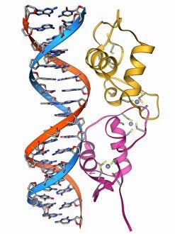
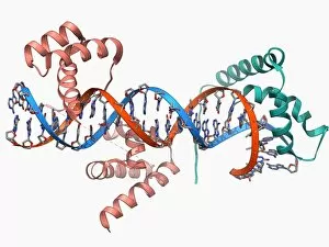
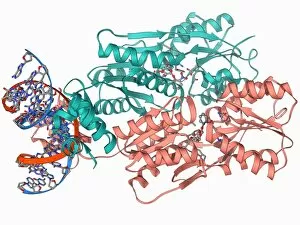
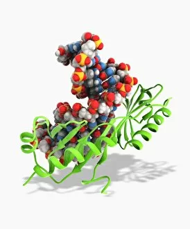
Background imageNucleic Acid Collection: Transcription factor and DNA molecule F006 / 9484

Transcription factor and DNA molecule F006 / 9484
Transcription factor and DNA molecule. Molecular model of glucocorticoid receptor (GR) transcription factor protein (pink and blue) complexed with a molecule of DNA (deoxyribonucleic acid)

Background imageNucleic Acid Collection: Androgen receptor, molecular model F006 / 9483

Androgen receptor, molecular model F006 / 9483
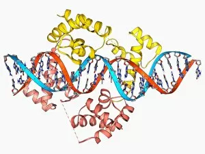
Androgen receptor. Molecular model of the DNA-binding region of an androgen receptor (pink and yellow) complexed with DNA (deoxyribonucleic acid, blue and red)
Background imageNucleic Acid Collection: RNA polymerase molecule F006 / 9475

RNA polymerase molecule F006 / 9475
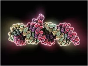
RNA polymerase. Molecular model of RNA polymerase (beige) transcribing a strand of mRNA (messenger ribonucleic acid, pink) from a DNA (deoxyribonucleic acid) template (red and blue)

Background imageNucleic Acid Collection: DNA transcription, molecular model F006 / 9424

DNA transcription, molecular model F006 / 9424
DNA transcription. Molecular model of the enzyme RNA polymerase II synthesising a mRNA (messenger ribonucleic acid) strand from a DNA (deoxyribonucleic acid) template

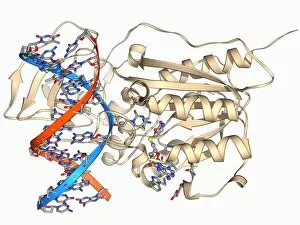
Background imageNucleic Acid Collection: Methyltransferase and DNA F006 / 9421

Methyltransferase and DNA F006 / 9421
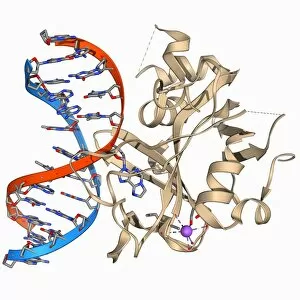
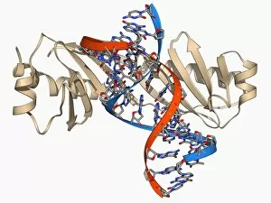
Methyltransferase and DNA. Molecular model of the enzyme HhaI methyltransferase (beige) complexed with a molecule of DNA (deoxyribonucleic acid, red and blue)

Background imageNucleic Acid Collection: Gene activator protein F006 / 9406

Gene activator protein F006 / 9406
Gene activator protein. Molecular model of catabolite gene activator protein (CAP, yellow) complexed with deoxyribonucleic acid (DNA, red and blue) and RNA polymerase (green and pink)

Background imageNucleic Acid Collection: Enzyme catalysing DNA recombination F006 / 9395

Enzyme catalysing DNA recombination F006 / 9395
Enzyme catalysing DNA recombination. Molecular model of the enzyme CRE (cyclization recombination) recombinase (green and purple) mediating the recombination of strands of DNA (deoxyribonucleic acid)

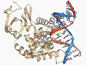
Background imageNucleic Acid Collection: DNA polymerase IV with DNA F006 / 9390

DNA polymerase IV with DNA F006 / 9390
polymerase IV with DNA. Molecular model of DNA polymerase IV (beige) complexed with a molecule of DNA (deoxyribonucleic acid, red and blue)

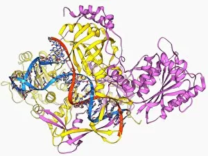
Background imageNucleic Acid Collection: Repair protein and DNA, molecular model F006 / 9382

Repair protein and DNA, molecular model F006 / 9382
Repair protein and DNA. Molecular model of the Ku heterodimer (yellow and pink) bound to a strand of DNA (deoxyribonucleic acid, red and blue) as part of the repair process

Background imageNucleic Acid Collection: DNA transcription, molecular model F006 / 9368

DNA transcription, molecular model F006 / 9368
DNA transcription. Molecular model of the enzyme RNA polymerase II synthesising a mRNA (messenger ribonucleic acid) strand from a DNA (deoxyribonucleic acid) template

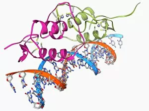
Background imageNucleic Acid Collection: Oestrogen receptor bound to DNA F006 / 9354

Oestrogen receptor bound to DNA F006 / 9354
Oestrogen receptor bound to DNA. Molecular model of the DNA-binding domain (green and pink) of the oestrogen receptor bound to a molecule of DNA (deoxyribonucleic acid, blue and orange)

Background imageNucleic Acid Collection: Ribozyme enzyme and RNA F006 / 9346

Ribozyme enzyme and RNA F006 / 9346
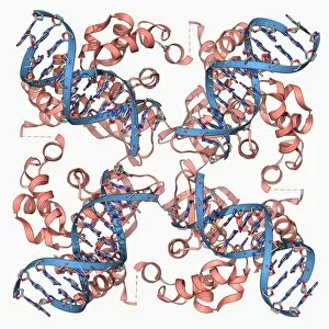
Ribozyme enzyme and RNA, molecular model. Ribozymes are RNA (ribonucleic acid) molecules that catalyse certain biochemical reactions

Background imageNucleic Acid Collection: Transcription factors bound to DNA F006 / 9349

Transcription factors bound to DNA F006 / 9349
Transcription factors bound to DNA. Molecular model of the Oct4 (pink) and Sox2 (green) transcription factors bound to a molecule of DNA (deoxyribonucleic acid, red and blue)

Background imageNucleic Acid Collection: DNA Holliday junction complex F006 / 9334

DNA Holliday junction complex F006 / 9334
DNA Holliday junction complex. Molecular model of the enzyme FLP recombinase in complex with a Holliday junction between homologous strands of DNA (deoxyribonucleic acid)

Background imageNucleic Acid Collection: Cre-Lox recombination, molecular model F006 / 9322

Cre-Lox recombination, molecular model F006 / 9322
Cre-Lox recombination. Molecular model of the enzyme CRE (cyclization recombination) recombinase (beige) mediating the recombination of strands of DNA (deoxyribonucleic acid, red and blue)

Background imageNucleic Acid Collection: Oxoguanine glycosylase complex F006 / 9318

Oxoguanine glycosylase complex F006 / 9318
Oxoguanine glycosylase complex. Computer model showing an 8-Oxoguanine glycosylase (OGG1) molecule (beige) bound to a section of DNA (deoxyribonucleic acid, red and blue)

Background imageNucleic Acid Collection: Restriction enzyme and DNA F006 / 9315

Restriction enzyme and DNA F006 / 9315
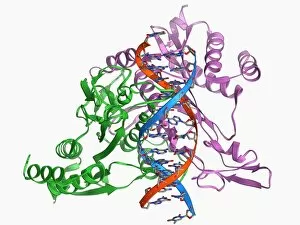
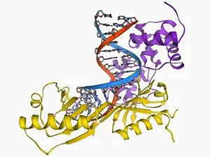
Restriction enzyme and DNA. Molecular model showing an EcoRI endonuclease enzyme (purple and green) bound to a DNA (deoxyribonucleic acid) molecule (red and blue)

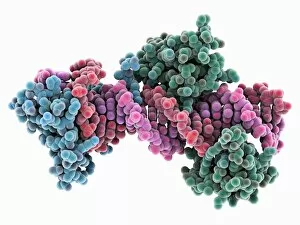
Background imageNucleic Acid Collection: LAC repressor bound to DNA F006 / 9309

LAC repressor bound to DNA F006 / 9309
LAC repressor bound to DNA. Molecular model of a LAC (lactose) repressor molecule (pink and turquoise) interacting with bacterial DNA (deoxyribonucleic acid, red and blue)

Background imageNucleic Acid Collection: Oxoguanine glycosylase complex F006 / 9307

Oxoguanine glycosylase complex F006 / 9307
Oxoguanine glycosylase complex. Computer model showing an 8-Oxoguanine glycosylase (OGG1) molecule (beige) bound to a section of DNA (deoxyribonucleic acid, red and blue)

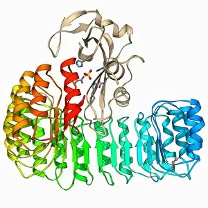
Background imageNucleic Acid Collection: Ribonuclease bound to inhibitor F006 / 9287

Ribonuclease bound to inhibitor F006 / 9287
Ribonuclease bound to inhibitor, molecular model. Ribonuclease (RNase) is a type of nuclease that catalyses the degradation of RNA (ribonucleic acid)

Background imageNucleic Acid Collection: DNA Holliday junction, molecular model F006 / 9285

DNA Holliday junction, molecular model F006 / 9285
DNA Holliday junction. Molecular model of a Holliday junction (centre) between homologous strands of DNA (deoxyribonucleic acid)

Background imageNucleic Acid Collection: Yeast DNA recognition, molecular model F006 / 9282

Yeast DNA recognition, molecular model F006 / 9282
Yeast DNA recognition. Computer model showing a GAL4 transcription activator protein bound to a yeast DNA (deoxyribonucleic acid) molecule (red and blue)

Background imageNucleic Acid Collection: DNA clamp complexed with DNA molecule F006 / 9274

DNA clamp complexed with DNA molecule F006 / 9274
DNA clamp complexed with DNA molecule. Molecular model showing a sliding DNA (deoxyribonucleic acid) clamp (beige) complexed with a molecule of DNA (red and blue)

Background imageNucleic Acid Collection: Gene activator protein F006 / 9269

Gene activator protein F006 / 9269
Gene activator protein. Molecular model of catabolite gene activator protein (CAP, pink and green) bound to a molecule of deoxyribonucleic acid (DNA, across top)

Background imageNucleic Acid Collection: TATA box-binding protein and DNA F006 / 9267

TATA box-binding protein and DNA F006 / 9267
TATA box-binding protein and DNA. Molecular model showing a TATA box-binding protein (TBP) complexed with a strand of DNA (deoxyribonucleic acid, red and blue)

Background imageNucleic Acid Collection: E coli Holliday junction complex F006 / 9261

E coli Holliday junction complex F006 / 9261
E. coli Holliday junction complex. Molecular model of a RuvA protein (red) in complex with a Holliday junction between homologous strands of DNA (deoxyribonucleic acid, blue) from an E

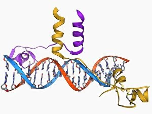
Background imageNucleic Acid Collection: Pit-1 transcription factor bound to DNA F006 / 9242

Pit-1 transcription factor bound to DNA F006 / 9242
Pit-1 transcription factor bound to DNA. Molecular model showing pituitary-specific positive transcription factor 1 (Pit-1) (yellow and pink) bound to a strand of DNA (deoxyribonucleic acid)

Background imageNucleic Acid Collection: TATA box-binding protein complex F006 / 9230

TATA box-binding protein complex F006 / 9230
TATA box-binding protein complex. Molecular model showing a yeast TATA box-binding protein (TBP) complexed with a strand of DNA (deoxyribonucleic acid, red and blue) and transcription factor IIB

Background imageNucleic Acid Collection: Type I topoisomerase bound to DNA F006 / 9221

Type I topoisomerase bound to DNA F006 / 9221
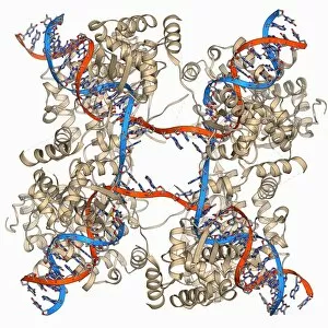
Type I topoisomerase bound to DNA. Molecular model showing a type I topoisomerase molecule (khaki) bound to a strand of DNA (deoxyribonucleic acid, red and blue)

Background imageNucleic Acid Collection: Type I topoisomerase bound to DNA F006 / 9220

Type I topoisomerase bound to DNA F006 / 9220
Type I topoisomerase bound to DNA. Molecular model showing a type I topoisomerase molecule (khaki) bound to a strand of DNA (deoxyribonucleic acid, red and blue)

Background imageNucleic Acid Collection: TATA box-binding protein complex C017 / 7089

TATA box-binding protein complex C017 / 7089
TATA box-binding protein complex. Molecular model showing a TATA box-binding protein (TBP, green) complexed with a strand of DNA (deoxyribonucleic acid, spheres) and transcription factor IIB

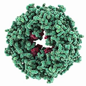
Background imageNucleic Acid Collection: Vault ribonucleoprotein molecule

Vault ribonucleoprotein molecule
Vault cytoplasmic ribonucleoprotein, molecular model. This molecule is made up from proteins and RNA (ribonucleic acid) and has a 39-fold symmetry

Background imageNucleic Acid Collection: Epstein-Barr virus protein and DNA

Epstein-Barr virus protein and DNA. Molecular model of the DNA-binding domain of a viral protein (pink-blue) bound to a lytic gene promoter element (viral strand of DNA, left)

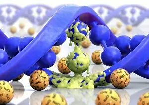
Background imageNucleic Acid Collection: Cystic fibrosis, conceptual artwork C015 / 5022

Cystic fibrosis, conceptual artwork C015 / 5022
Cystic fibrosis, conceptual computer artwork. Molecule of DNA (deoxyribonucleic acid, blue) partly covered in mucus (yellow) surrounded by salt (NaCl) molecules

Background imageNucleic Acid Collection: DNA molecule, artwork C016 / 8892

DNA molecule, artwork C016 / 8892
DNA molecule. Computer artwork showing a double stranded DNA (deoxyribonucleic acid) molecule. DNA is composed of two strands twisted into a double helix

Background imageNucleic Acid Collection: Biotin-binding RNA molecule

Biotin-binding RNA molecule
Biotin-binding RNA (ribonucleic acid), molecular model. This RNA molecule is a pseudoknot, formed from stem-loop structures. It binds to the vitamin B7 (biotin)

Background imageNucleic Acid Collection: DNA repair enzyme, molecular model

DNA repair enzyme, molecular model
DNA repair enzyme. Molecular model of a recG molecule (purple) complexed with a three-way DNA (deoxyribonucleic acid, pink, green and yellow) junction

Background imageNucleic Acid Collection: Genomic HIV-RNA duplex

Genomic HIV-RNA duplex, molecular model. This structure shows the dimerization initiation site of genomic HIV-1 with RNA (ribonucleic acid)

Background imageNucleic Acid Collection: Ebola matrix protein molecule

Ebola matrix protein molecule
Ebola matrix protein. Molecular model of the Ebola virus matrix protein VP40 (green) bound to RNA (ribonucleic acid, red)

Background imageNucleic Acid Collection: SelB elongation factor bound to RNA

SelB elongation factor bound to RNA. Molecular model of the SelB elongation factor bound to an mRNA (messenger ribonucleic acid) hairpin formed by the selenocysteine insertion sequence (SECIS)

Background imageNucleic Acid Collection: HU DNA binding protein molecule

HU DNA binding protein molecule
HU DNA binding protein. Molecular model of the HU protein (orange and blue) bound to a molecule of DNA (deoxyribonucleic acid)

Background imageNucleic Acid Collection: Cre-Lox recombination, molecular model

Cre-Lox recombination, molecular model
Cre-Lox recombination. Molecular model of the enzyme CRE (cyclization recombination) recombinase (blue) mediating the recombination of strands of DNA (deoxyribonucleic acid, pink and yellow)

Background imageNucleic Acid Collection: Oct and Sox transcription factors

Oct and Sox transcription factors. Molecular model of Sox2 (blue) and Oct4 (green) transcription factors bound to a molecule of DNA (deoxyribonucleic acid, pink and red)

Background imageNucleic Acid Collection: DNA bundle on silicon nanopillars, SEM

DNA bundle on silicon nanopillars, SEM
DNA bundle on silicon nanopillars. Scanning electron micrograph (SEM) of a DNA (deoxyribonucleic acid) bundle and silicon nanopillars used to obtain the first high-contrast direct images of DNA

Background imageNucleic Acid Collection: MyoD muscle protein-DNA complex

MyoD muscle protein-DNA complex. Molecular model of the MyoD muscle protein (green and blue) bound to a strand of DNA (deoxyribonucleic acid, orange and green)

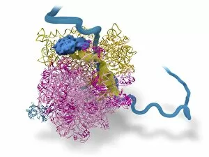
Background imageNucleic Acid Collection: Bacterial ribosome and protein synthesis

Bacterial ribosome and protein synthesis. Molecular model showing a bacterial ribosome reading an mRNA (messenger ribonucleic acid) strand (blue) and synthesising a protein

Background imageNucleic Acid Collection: DNA repair protein AlkB with DNA C016 / 0547

DNA repair protein AlkB with DNA C016 / 0547
DNA repair protein AlkB with DNA. Molecular model of the DNA (deoxyribonucleic acid) repair protein AlkB (blue) bound to a strand of double-stranded DNA (ds-DNA, pink and yellow)

Background imageNucleic Acid Collection: RNA silencing molecule C015 / 7697

RNA silencing molecule C015 / 7697
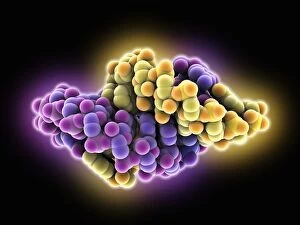
RNA silencing. Molecular model of a synthetic double stranded ribonucleic acid (RNA) molecule. RNA is the intermediate molecule between DNA (deoxyribonucleic acid) and its protein products


For sale as Licensed Images

Choose your image, Select your licence and Download the media

Nucleic acids, the building blocks of life, are intricately woven strands of genetic information that hold the key to our existence. In this captivating journey into their world, we explore the wonders they unveil. A mesmerizing sight awaits as we gaze upon a double-stranded RNA molecule, its elegant structure resembling a delicate dance of intertwined ribbons. Next, a computer model unveils the intricate beauty of a DNA molecule, showcasing its unique helical shape and revealing the blueprint for life itself. Moving deeper into this microscopic realm, we encounter a DNA nucleosome in all its glory - a molecular masterpiece where DNA elegantly wraps around histone proteins like an artistic sculpture. An artwork depicting another DNA molecule captures our imagination with vibrant colors and abstract patterns that symbolize the complexity hidden within. Zinc fingers bound to a DNA strand create an enchanting spectacle as they delicately interact with each other like tiny keys unlocking genetic secrets. The iconic image of the DNA Double Helix with Autoradiograph reminds us of Rosalind Franklin's pioneering work in unraveling nature's code. Diving further into this fascinating world, we come across Z-DNA tetramer molecules standing tall like architectural marvels - their distinct zigzag pattern hinting at alternative possibilities within our genetic makeup. A molecular model showcases an RNA-editing enzyme poised for action; it is through these enzymes that our genes can be fine-tuned and modified. The journey continues with yet another glimpse at the awe-inspiring simplicity and complexity coexisting within a single DNA molecule. Ribonuclease gracefully interacts with an RNA/DNA hybrid - highlighting how these molecules intertwine to carry out essential cellular functions. Intriguingly conceptualized artistry takes center stage as creation unfolds before our eyes - reminding us that they are not just passive observers but active participants in shaping life's tapestry. Finally, an illustration encapsulates the essence of nucleic acid, capturing the essence of their importance in a single image.