
Nucleic Acid Collection (#8)
Nucleic acids, the building blocks of life, are intricately woven strands of genetic information that hold the key to our existence


For sale as Licensed Images
Choose your image, Select your licence and Download the media
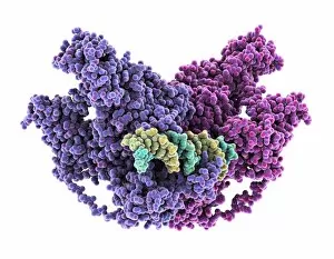
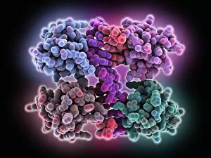
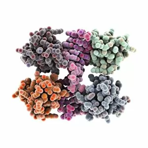
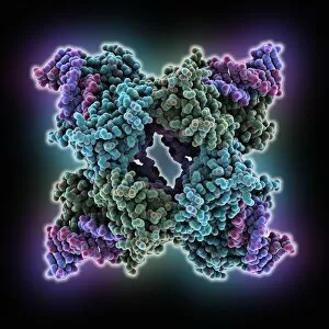
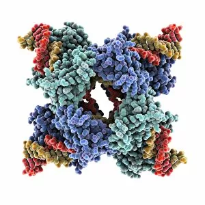
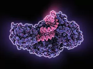
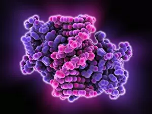
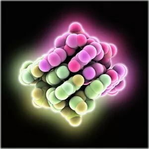
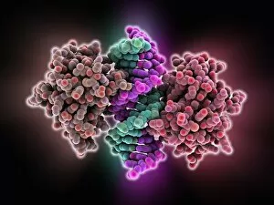
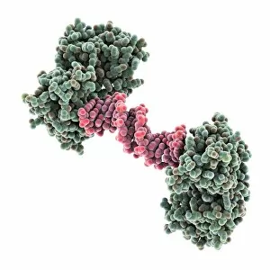
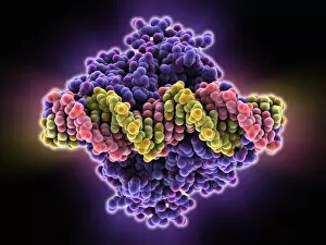
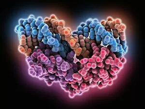
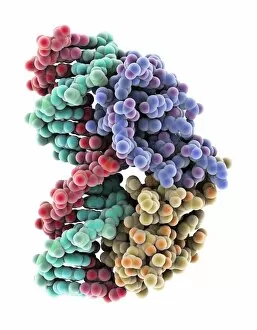
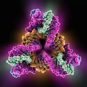
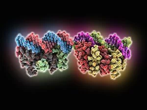
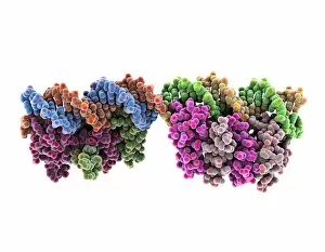
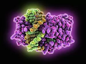
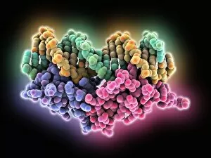
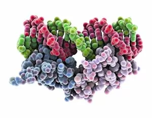
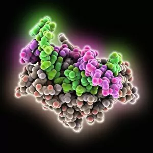
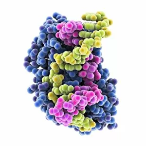
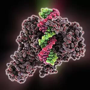
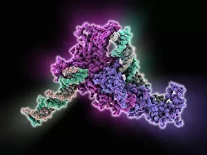
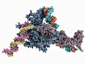
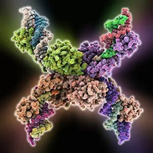
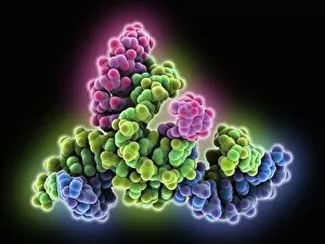
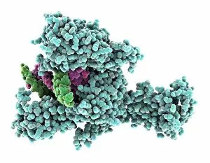
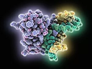
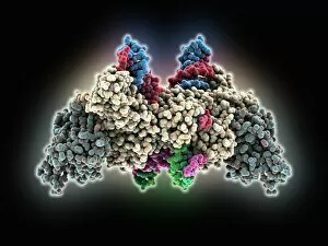
Nucleic acids, the building blocks of life, are intricately woven strands of genetic information that hold the key to our existence. In this captivating journey into their world, we explore the wonders they unveil. A mesmerizing sight awaits as we gaze upon a double-stranded RNA molecule, its elegant structure resembling a delicate dance of intertwined ribbons. Next, a computer model unveils the intricate beauty of a DNA molecule, showcasing its unique helical shape and revealing the blueprint for life itself. Moving deeper into this microscopic realm, we encounter a DNA nucleosome in all its glory - a molecular masterpiece where DNA elegantly wraps around histone proteins like an artistic sculpture. An artwork depicting another DNA molecule captures our imagination with vibrant colors and abstract patterns that symbolize the complexity hidden within. Zinc fingers bound to a DNA strand create an enchanting spectacle as they delicately interact with each other like tiny keys unlocking genetic secrets. The iconic image of the DNA Double Helix with Autoradiograph reminds us of Rosalind Franklin's pioneering work in unraveling nature's code. Diving further into this fascinating world, we come across Z-DNA tetramer molecules standing tall like architectural marvels - their distinct zigzag pattern hinting at alternative possibilities within our genetic makeup. A molecular model showcases an RNA-editing enzyme poised for action; it is through these enzymes that our genes can be fine-tuned and modified. The journey continues with yet another glimpse at the awe-inspiring simplicity and complexity coexisting within a single DNA molecule. Ribonuclease gracefully interacts with an RNA/DNA hybrid - highlighting how these molecules intertwine to carry out essential cellular functions. Intriguingly conceptualized artistry takes center stage as creation unfolds before our eyes - reminding us that they are not just passive observers but active participants in shaping life's tapestry. Finally, an illustration encapsulates the essence of nucleic acid, capturing the essence of their importance in a single image.