Nucleus Collection (page 2)

"The Nucleus: Unveiling the Mysteries of Life's Command Center" Exploring the intricate world within our cerebellum tissue

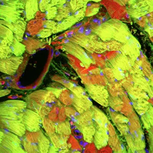
Background imageNucleus Collection: Heart muscle, confocal light micrograph

Heart muscle, confocal light micrograph
Heart muscle. Confocal light micrograph of a section through cardiac (heart) muscle. Cardiac muscle consists of branching elongated muscle cells

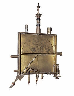
Background imageNucleus Collection: Model of cyclotron, particle accelerator, invented by Ernest Orlando Lawrence (1901-1958)

Model of cyclotron, particle accelerator, invented by Ernest Orlando Lawrence (1901-1958)
AIS5378122 Model of cyclotron, particle accelerator, invented by Ernest Orlando Lawrence (1901-1958) and Milton Stanley Livingston (1905-1986) in 1931 (metal object); Science Museum, London

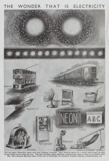
Background imageNucleus Collection: The wonder that is electricity (litho)

The wonder that is electricity (litho)
8676245 The wonder that is electricity (litho) by English School, (20th century); Private Collection; (add.info.: The wonder that is electricity)

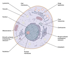
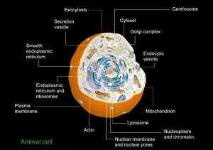
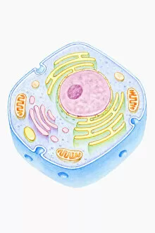
Background imageNucleus Collection: Animal cell structure

Animal cell structure. Artwork of a section through an animal cell. At lower centre is the nucleus, which contains the cells genetic information in the form of DNA (deoxyribonucleic acid)

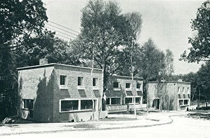
Background imageNucleus Collection: Group Of Modernist Houses, Haywards Heath

Group Of Modernist Houses, Haywards Heath
A photograph of a group of modernist houses forming the nucleus of a housing estate in Haywards Heath, Sussex, designed in 1936 by architect Berthold Romanovich Lubetkin and Tecton. Date: circa 1937

Background imageNucleus Collection: Warty crab and flat rock crab

Warty crab and flat rock crab
Warty crab or yellow crab, Eriphia verrucosa 1, and flat rock crab, Percnon planissimum 2. Erifia fronte spinosa, Plagusia clavimana

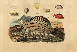
Background imageNucleus Collection: Tiger cowrie, wrinkled cowrie, money cowrie, etc

Tiger cowrie, wrinkled cowrie, money cowrie, etc
Tiger cowrie, Cypraea tigris 1, wrinkled cowrie, Nucleolaria nucleus 2, money cowrie, Monetaria moneta 3, and Ellatrivia merces 4, Porcelaines

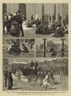
Background imageNucleus Collection: Egypt after the War, Lord Dufferin at Cairo (engraving)

Egypt after the War, Lord Dufferin at Cairo (engraving)
1061236 Egypt after the War, Lord Dufferin at Cairo (engraving) by English School, (19th century); Private Collection; (add.info.: Egypt after the War, Lord Dufferin at Cairo)

Background imageNucleus Collection: The War in South Africa (litho)

The War in South Africa (litho)
1599684 The War in South Africa (litho) by Stewart, Frank Algenon (1877-1945); Private Collection; (add.info.: The War in South Africa. Illustration for The Illustrated London News, 26 May 1900)

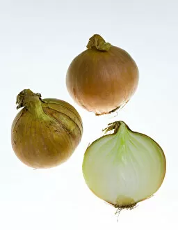
Background imageNucleus Collection: EyeUbiquitous_20104516

EyeUbiquitous_20104516
Food, Vegetables, Onions, Three ripe harvested onion bulbs or Allium against a white background with one sliced open to show structure of layers

Background imageNucleus Collection: Ian Carr, Nucleus, Ronnie Scotts, Soho London, May 1991. Creator: Brian O Connor

Ian Carr, Nucleus, Ronnie Scotts, Soho London, May 1991. Creator: Brian O Connor
Ian Carr, Nucleus, Ronnie Scott s, Soho, London, May 1991

Background imageNucleus Collection: Illustration of cell nucleus containing cell cytoplasm, mitochondria units, DNA, and chromosome

Illustration of cell nucleus containing cell cytoplasm, mitochondria units, DNA, and chromosome

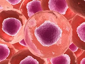
Background imageNucleus Collection: Microscopic view of animal cell

Microscopic view of animal cell

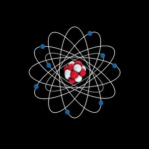
Background imageNucleus Collection: Oxygen atomic structure, artwork

Oxygen atomic structure, artwork
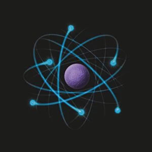
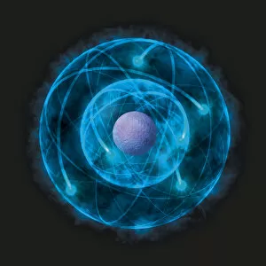
Oxygen atomic structure. Computer artwork showing the structure of an oxygen atom. Each oxygen atom contains 8 electrons (blue) orbiting the atomic nucleus (centre)

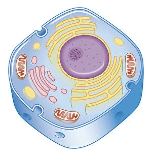
Background imageNucleus Collection: Animal cell anatomy, diagram

Animal cell anatomy, diagram
Animal cell anatomy. Diagram showing the internal and external anatomy of an animal cell

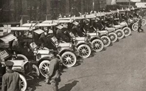
Background imageNucleus Collection: A row of taxi cabs waiting to take Automobile Association offiers

A row of taxi cabs waiting to take Automobile Association offiers and patrols to the White City to form the nucleus of a new cyclist battalion, the Army Cyclist Corps, at the start of WWI

Background imageNucleus Collection: Illustration of Rutherford model of the atom, consisting of nucleus and negatively charged electrons

Illustration of Rutherford model of the atom, consisting of nucleus and negatively charged electrons

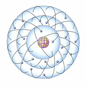
Background imageNucleus Collection: Illustration of atom with nucleus of protons and neurons, based on the Bohr model

Illustration of atom with nucleus of protons and neurons, based on the Bohr model

Background imageNucleus Collection: Illustration of neutron hitting Uranium-235 nucleus, nucleus becoming unstable and splitting

Illustration of neutron hitting Uranium-235 nucleus, nucleus becoming unstable and splitting, releasing energy and neutrons (nuclear fission)

Background imageNucleus Collection: Retina with macular degeneration

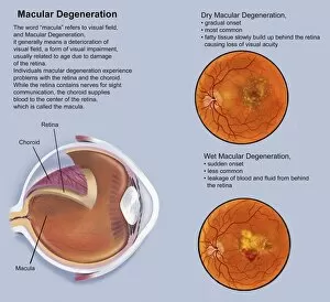
Retina with macular degeneration

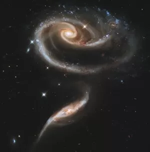
Background imageNucleus Collection: Arp 273 interacting galaxies in Andromeda

Arp 273 interacting galaxies in Andromeda. The larger of the two galaxies, UGC 1810, shows a large spiral galaxy with a disk that is distorted into a rose-like shape by the gravitational tidal pull

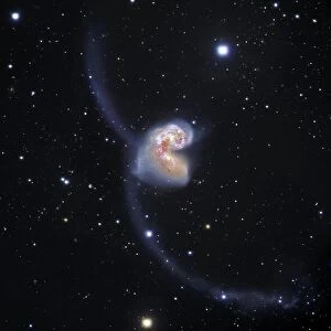
Background imageNucleus Collection: Antennae colliding galaxies C017 / 3745

Antennae colliding galaxies C017 / 3745
Antennae colliding galaxies, combined optical image. The Antennae (NGC 4038 and NGC 4039) are formed of two galaxies colliding due to mutual gravitational attraction

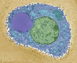
Background imageNucleus Collection: Pancreatic acinar cell

Pancreatic acinar cell. Transmission electron micrograph (TEM) of a section through an enzyme-secreting acinar cell in the human pancreas, showing part of the nucleus (round, far left)

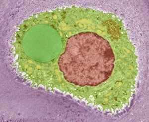
Background imageNucleus Collection: Cartilage cell, TEM C014 / 1433

Cartilage cell, TEM C014 / 1433
Cartilage cell. Transmission electron micrograph (TEM) of a section through a chondrocyte cell from hyaline cartilage of the trachea (windpipe)

Background imageNucleus Collection: Cartilage cell, TEM C014 / 1434

Cartilage cell, TEM C014 / 1434
Cartilage cell. Transmission electron micrograph (TEM) of a section through a chondrocyte cell from hyaline cartilage of the trachea (windpipe)

Background imageNucleus Collection: Beryllium, atomic model

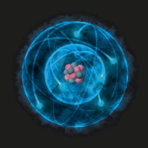
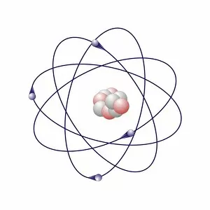
Beryllium, atomic model. Beryllium has five neutrons (white) and four protons (pink) in its nucleus (centre). The atom also has four electron (blue) orbiting the nucleus

Background imageNucleus Collection: Helium, atomic model

Helium, atomic model
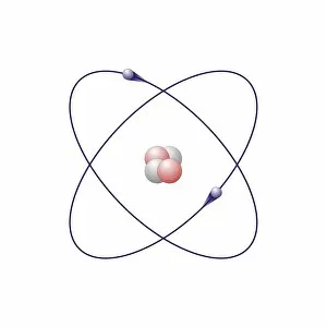
Heium, atomic model. Helium has two neutrons (white) and two protons (pink) in its nucleus (centre). The atom also has two electron (blue) orbiting the nucleus

Background imageNucleus Collection: Boron, atomic model

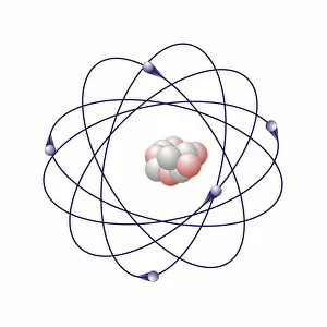
Boron, atomic model. Boron has six neutrons (white) and five protons (pink) in its nucleus (centre). The atom also has five electron (blue) orbiting the nucleus

Background imageNucleus Collection: English Cathedrals - Gloucester - the Nave

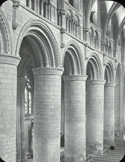
English Cathedrals - Gloucester - the Nave - Ornamental stone arches between the columns. Part of Box 144 Boswell Collection - English Cathedrals

Background imageNucleus Collection: Spain. Citadel of Roses (Rhode). Roman ruins

Spain. Citadel of Roses (Rhode). Roman ruins
Spain. Citadel of Roses (Rhode). Excavations of a urban nucleus of the Low Roman Empire. 3rd-2nd centuries BC

Background imageNucleus Collection: Pre-romanesque Church of Saint Peter. Terrassa. Spain

Pre-romanesque Church of Saint Peter. Terrassa. Spain
Pre-romanesque Church of Saint Peter. View of the trefoil apse. 9th-10th centuries. Terrassa. Catalonia. Spain

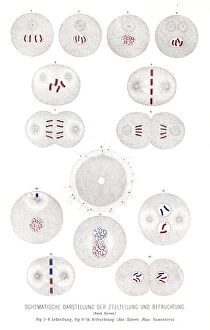
Background imageNucleus Collection: Cell division and fertilization anatomy engraving 1857

Cell division and fertilization anatomy engraving 1857
Rank, johannes - The human being. 1 - 1894

Background imageNucleus Collection: Cross section illustration of human cell

Cross section illustration of human cell

Background imageNucleus Collection: Illustration of human nerve cell cross section showing dendrite, soma, axon, nucleus

Illustration of human nerve cell cross section showing dendrite, soma, axon, nucleus, nodes and myelin sheath

Background imageNucleus Collection: Illustration showing relative atomic mass of nucleus of boron-11 containing five protons

Illustration showing relative atomic mass of nucleus of boron-11 containing five protons and six neutrons

Background imageNucleus Collection: Electrons spinning around nucleus of an atom

Electrons spinning around nucleus of an atom

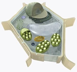
Background imageNucleus Collection: Illustration of the structure of a plant cell, including nucleus, nucleolus, ribosome, endoplasmatic

Illustration of the structure of a plant cell, including nucleus, nucleolus, ribosome, endoplasmatic

Background imageNucleus Collection: Illustration of how atoms emit light, based on Bohr model

Illustration of how atoms emit light, based on Bohr model

Background imageNucleus Collection: Illustration of Bohr model of the atom

Illustration of Bohr model of the atom

Background imageNucleus Collection: Digital cross section illustration of impulses passing from nearer cochlea nucleus to lateral

Digital cross section illustration of impulses passing from nearer cochlea nucleus to lateral superior olive

Background imageNucleus Collection: Illustration of nucleus of Hydrogen-2 and nucleus of Hydrogen-3 fusing

Illustration of nucleus of Hydrogen-2 and nucleus of Hydrogen-3 fusing and forming nucleus of Helium-4, expelling a neutron (nuclear fusion)

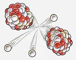
Background imageNucleus Collection: Illustration of split atoms and neutrons

Illustration of split atoms and neutrons

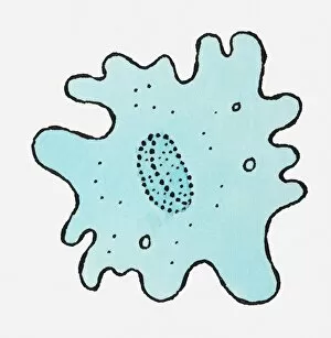
Background imageNucleus Collection: Illustration of blue Amoeba

Illustration of blue Amoeba

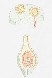
Background imageNucleus Collection: Illustration of fertilization of flower, pollen grain containing male nuclei on stigma

Illustration of fertilization of flower, pollen grain containing male nuclei on stigma, male nucleus travelling down pollen tube, female nuclei inside ovule

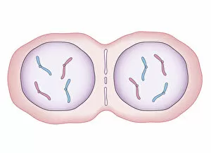
Background imageNucleus Collection: Cross section biomedical illustration of mitosis where nucleus membrane form around each set of

Cross section biomedical illustration of mitosis where nucleus membrane form around each set of chromosomes and the cell begins to divide in two

Background imageNucleus Collection: Cross section biomedical illustration of mitosis with new cells form

Cross section biomedical illustration of mitosis with new cells form, each having a central nucleus containing identical set of chromosomes

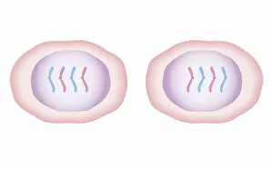
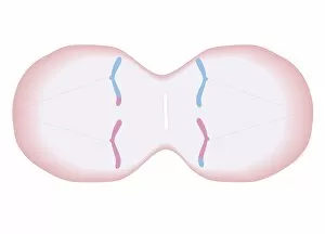
Background imageNucleus Collection: Cross section biomedical illustration of meiosis with duplicated chromosomes lined up

Cross section biomedical illustration of meiosis with duplicated chromosomes lined up and more threads attach, pulling the duplicated chromosomes apart to form two single chromosomes

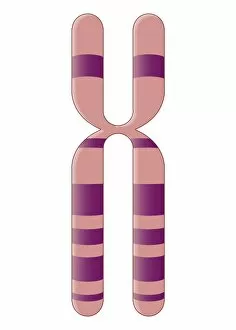
Background imageNucleus Collection: Biomedical illustration of X Chromosome

Biomedical illustration of X Chromosome


For sale as Licensed Images

Choose your image, Select your licence and Download the media

"The Nucleus: Unveiling the Mysteries of Life's Command Center" Exploring the intricate world within our cerebellum tissue, where the nucleus orchestrates every movement and balance. A mesmerizing light micrograph reveals the nucleus at the heart of M51 Whirlpool Galaxy, a cosmic dance guided by its gravitational pull. Witnessing life's beginning through a budding yeast cell, as its nucleus holds the blueprint for growth and reproduction. Delving into the secrets of communication with a nerve cell captured in stunning detail through SEM imaging, showcasing its complex nucleus. Behold the majestic Sombrero Galaxy (Messier 104), where billions of stars revolve around their central nucleus like celestial dancers in perfect harmony. The hippocampus brain tissue unveils memories etched within each neuron's nucleus, shaping who we are and what we remember. Peering into HeLa cells under a light microscope (C017/8299), revealing their remarkable nuclei that have revolutionized medical research. An artistic representation takes us deep into Medulla Oblongata's core—the vital center controlling essential bodily functions—where nuclei reign supreme. Captivating artwork depicts nuclear fission—a powerful force harnessed to generate energy while reminding us of its immense potential and responsibility. Marvel at NGC 4258 spiral galaxy's galactic light show, where vibrant colors illuminate star clusters revolving around their radiant nuclear core. Human cells come alive under scrutiny as their nuclei hold genetic information that shapes our unique characteristics and existence itself. Exploring kidney tubules in section unravels an intricate network governed by countless tiny nuclei working harmoniously to maintain our body’s equilibrium. Intricate yet awe-inspiring, these glimpses into various realms remind us that no matter how vast or minuscule, from galaxies to microscopic cells - all bow to the commanding presence of the nucleus.