Nucleus Collection (#9)

"The Nucleus: Unveiling the Mysteries of Life's Command Center" Exploring the intricate world within our cerebellum tissue

Background imageNucleus Collection: Collection of frogspawn

Collection of frogspawn

Background imageNucleus Collection: Stochastic gene expression, illustration C018 / 0906

Stochastic gene expression, illustration C018 / 0906
Stochastic gene expression, illustration. Every cell in an organism contains every single gene that makes up the organisms genome. However, they are not all active (expressed) in each cell

Background imageNucleus Collection: Artwork of water-rich asteroid

Artwork of water-rich asteroid
Artwork showing an asteroid breaking up under the gravitational influence of a nearby white dwarf star. In looking at debris discs around nearby stars

Background imageNucleus Collection: Skeletal muscle fibres, light micrograph

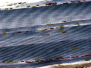
Skeletal muscle fibres, light micrograph
Skeletal muscle fibres. Light micrograph of skeletal muscle fibres showing the characteristic banding. Cell nuclei are yellow and red blood cells, in capillaries in the endomycium, are red

Background imageNucleus Collection: Atom, artwork

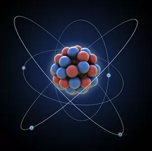
Atom, artwork
Atom, computer artwork

Background imageNucleus Collection: Particles in forcefield, artwork

Particles in forcefield, artwork
Conceptual computer artwork depicting particles in a force field

Background imageNucleus Collection: Artwork of nuclear fusion reaction

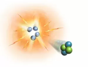
Artwork of nuclear fusion reaction
Conceptual image representing the process of nuclear fusion, specifically the creation of helium from hydrogen. Four protons (hydrogen nuclei) are combining on the left

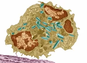
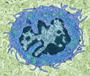
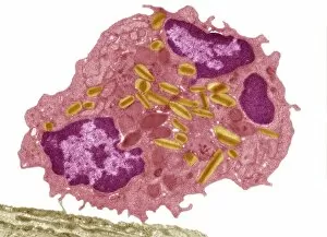
Background imageNucleus Collection: Eosinophil white blood cell, TEM C014 / 1438

Eosinophil white blood cell, TEM C014 / 1438
Eosinophil white blood cell. Transmission electron micrograph (TEM) of a section through an eosinophil. Eosinophils, like all white blood cells, are part of the bodys immune system

Background imageNucleus Collection: Comet 65P-Gunn, infrared image

Comet 65P-Gunn, infrared image. This comet is a short-period one (6.79 years) that orbits the Sun inside the main asteroid belt between the orbits of the planets Mars and Jupiter

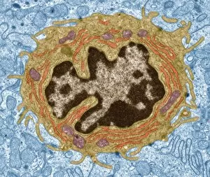
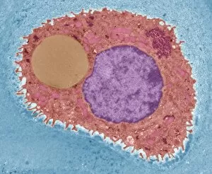
Background imageNucleus Collection: Intestinal gland cell, TEM C014 / 1441

Intestinal gland cell, TEM C014 / 1441
Intestinal gland cell. Transmission electron micrograph (TEM) of a section through an undifferentiated cell from the intestinal tract, showing extensive rough endoplasmic reticulum (ER)

Background imageNucleus Collection: Praseodymium, atomic structure

Praseodymium, atomic structure
Bismuth (Bi). Diagram of the nuclear composition, electron configuration, chemical data, and valence orbitals of an atom of bismuth-209 (atomic number: 83), the most common isotope of this element

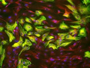
Background imageNucleus Collection: Astrocyte brain cells, light micrograph

Astrocyte brain cells, light micrograph
Astrocyte brain cells. Fluorescence light micrograph of primary astrocyte cells from the brain of a mouse. Astrocytes have numerous branches of connective tissue that provide support

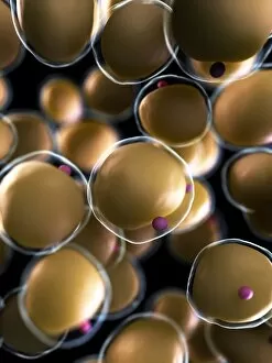
Background imageNucleus Collection: Human fat cells, artwork F006 / 2275

Human fat cells, artwork F006 / 2275
Human fat cells, computer artwork

Background imageNucleus Collection: Human fat cells, artwork F006 / 2274

Human fat cells, artwork F006 / 2274
Human fat cells, computer artwork

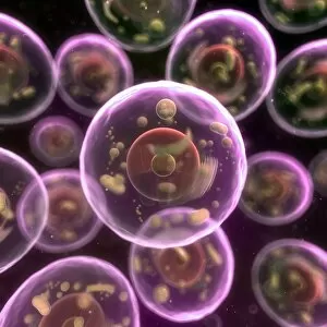
Background imageNucleus Collection: Animal cells, artwork F006 / 2225

Animal cells, artwork F006 / 2225
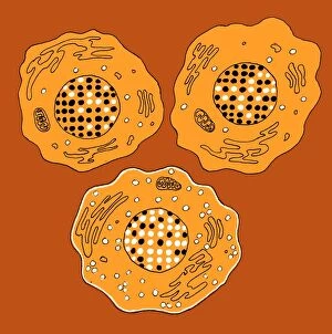
Animal cells, computer artwork

Background imageNucleus Collection: Animal cells, artwork F006 / 2224

Animal cells, artwork F006 / 2224
Animal cells, computer artwork

Background imageNucleus Collection: Animal cells, artwork F006 / 2223

Animal cells, artwork F006 / 2223
Animal cells, computer artwork

Background imageNucleus Collection: Animal cells, artwork F006 / 2222

Animal cells, artwork F006 / 2222
Animal cells, computer artwork

Background imageNucleus Collection: Intestinal gland cell, TEM C014 / 1440

Intestinal gland cell, TEM C014 / 1440
Intestinal gland cell. Transmission electron micrograph (TEM) of a section through an undifferentiated cell from the intestinal tract, showing extensive rough endoplasmic reticulum (ER)

Background imageNucleus Collection: Cartilage cell, TEM C014 / 1432

Cartilage cell, TEM C014 / 1432
Cartilage cell. Transmission electron micrograph (TEM) of a section through a chondrocyte cell from hyaline cartilage of the trachea (windpipe)

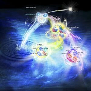
Background imageNucleus Collection: Structure of matter, artwork C018 / 0948

Structure of matter, artwork C018 / 0948
Structure of matter. Computer artwork representing the Standard Model of particle physics. Shown here is a molecule of water (top centre)

Background imageNucleus Collection: Phosphorus, atomic structure C018 / 3696

Phosphorus, atomic structure C018 / 3696
Argon (Ar). Diagram of the nuclear composition, electron configuration, chemical data, and valence orbitals of an atom of argon-40 (atomic number: 18)

Background imageNucleus Collection: Nucleus and endoplasmic reticulum F006 / 9196

Nucleus and endoplasmic reticulum F006 / 9196
Computer artwork showing part of a human or eukaryotic cell. In the middle the nucleus which has a membrane with nuclear pores. Inside the nucleus is the DNA

Background imageNucleus Collection: Animal cell, artwork F006 / 9019

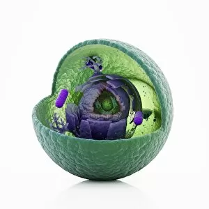
Animal cell, artwork F006 / 9019
Animal cell, cut-away computer artwork

Background imageNucleus Collection: Atom, artwork F006 / 8760

Atom, artwork F006 / 8760
Atom. Schematic diagram of an atom

Background imageNucleus Collection: Cell division, artwork F006 / 9891

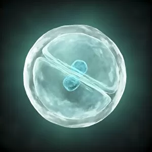
Cell division, artwork F006 / 9891
Cell division, computer artwork

Background imageNucleus Collection: Cell division, artwork F006 / 9892

Cell division, artwork F006 / 9892
Cell division, computer artwork

Background imageNucleus Collection: Headache, conceptual artwork F006 / 8693

Headache, conceptual artwork F006 / 8693
Headache, conceptual computer artwork

Background imageNucleus Collection: Plant cell, artwork F006 / 9876

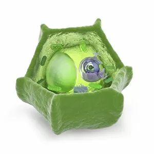
Plant cell, artwork F006 / 9876
Plant cell, computer artwork

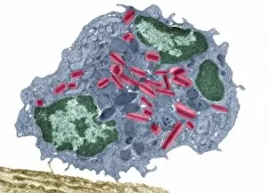
Background imageNucleus Collection: Eosinophil white blood cell, TEM C014 / 1437

Eosinophil white blood cell, TEM C014 / 1437
Eosinophil white blood cell. Transmission electron micrograph (TEM) of a section through an eosinophil. Eosinophils, like all white blood cells, are part of the bodys immune system

Background imageNucleus Collection: Packaged DNA, conceptual artwork F006 / 9861

Packaged DNA, conceptual artwork F006 / 9861
Packaged DNA, conceptual computer artwork

Background imageNucleus Collection: Packaged DNA, conceptual artwork F006 / 9853

Packaged DNA, conceptual artwork F006 / 9853
Packaged DNA, conceptual computer artwork

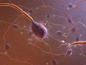
Background imageNucleus Collection: Nerve synapse, computer artwork C017 / 3429

Nerve synapse, computer artwork C017 / 3429
Nerve synapse. Conceptual computer artwork of a synapse between two nerve cells or neurons. Neurons are responsible for passing information around the central nervous system (CNS)

Background imageNucleus Collection: Brain nerve cells, TEM C014 / 0357

Brain nerve cells, TEM C014 / 0357
Brain nerve cells. Transmission electron micrograph (TEM) of a section through brain tissue from the cerebral cortex, showing numerous neurons (nerve cells) surrounded by axons and dendrites

Background imageNucleus Collection: Eosinophil white blood cell, TEM C014 / 1439

Eosinophil white blood cell, TEM C014 / 1439
Eosinophil white blood cell. Transmission electron micrograph (TEM) of a section through an eosinophil. Eosinophils, like all white blood cells, are part of the bodys immune system

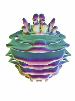
Background imageNucleus Collection: Golgi apparatus, artwork F006 / 9211

Golgi apparatus, artwork F006 / 9211
Computer artwork of the Golgi apparatus of the human cell. This organelle functions as a central delivery system for the cell

Background imageNucleus Collection: Nucleus and endoplasmic reticulum F006 / 9201

Nucleus and endoplasmic reticulum F006 / 9201
Computer artwork showing part of a human or eukaryotic cell. In the middle the nucleus which has a membrane with nuclear pores. Inside the nucleus is the DNA

Background imageNucleus Collection: Golgi apparatus, artwork F006 / 9199

Golgi apparatus, artwork F006 / 9199
Computer artwork of the Golgi apparatus of the human cell. This organelle functions as a central delivery system for the cell

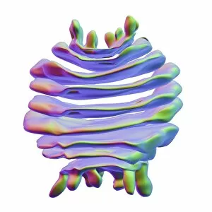
Background imageNucleus Collection: Golgi apparatus, artwork F006 / 9189

Golgi apparatus, artwork F006 / 9189
Computer artwork of the Golgi apparatus of the human cell. This organelle functions as a central delivery system for the cell

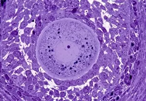
Background imageNucleus Collection: Oocyte, light micrograph

Oocyte, light micrograph
Oocyte. Light micrograph of a section through an oocyte within a early antral follicle in an ovary. Oocytes are immature ova, or egg cells

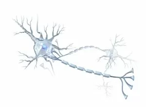
Background imageNucleus Collection: Nerve cells, artwork C015 / 4265

Nerve cells, artwork C015 / 4265
Nerve cells. Computer artwork of two nerve cells or neurons. Neurons are responsible for passing information around the central nervous system (CNS) and from the CNS to the rest of the body

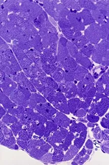
Background imageNucleus Collection: Cardiac muscle, light micrograph

Cardiac muscle, light micrograph
Cardiac muscle. Light micrograph of a section through muscle tissue from a heart, showing the cardiomyocyte cells. Heart muscle cells show central nuclei but the dominant feature is the abundance of

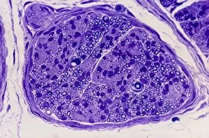
Background imageNucleus Collection: Peripheral nerve, light micrograph

Peripheral nerve, light micrograph
Peripheral nerve. Light micrograph of a section through a peripheral nerve. This is a mixed nerve with myelinated axons (dark blue circles)

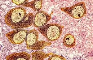
Background imageNucleus Collection: Nerve ganglion, light micrograph

Nerve ganglion, light micrograph
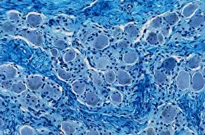
Nerve ganglion. Light micrograph of a section through a dorsal (sensory) spinal root ganglion associated with a sensory nerve root of the spinal cord. Sensory information from peripheral sites e.g

Background imageNucleus Collection: Myelinated nerve, light micrograph

Myelinated nerve, light micrograph
Myelinated nerve. Light micrograph of a section through a peripheral myelinated nerve, showing many individual axons each covered with deep blue-staining myelin sheaths

Background imageNucleus Collection: Fertilisation, artwork C016 / 7516

Fertilisation, artwork C016 / 7516
Fertilisation. Cutaway computer artwork of a sperm cell (spermatozoa) penetrating an egg (large). The nucleus of the sperm cell will fuse with the egg and its DNA (deoxyribonucleic acid)

Background imageNucleus Collection: DNA packaging, artwork C016 / 7517

DNA packaging, artwork C016 / 7517
DNA packaging. Computer artwork showing how DNA (deoxyribonucleic acid) is packaged within cells. Two DNA strands, consisting of a sugar-phosphate backbone attached to nucleotide bases

Background imageNucleus Collection: Corpus luteum, TEM

Corpus luteum, TEM
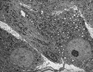
Corpus luteum. Transmission electron micrograph (TEM) of a section through the corpus luteum of an ovary, showing several luteal cells


For sale as Licensed Images

Choose your image, Select your licence and Download the media

"The Nucleus: Unveiling the Mysteries of Life's Command Center" Exploring the intricate world within our cerebellum tissue, where the nucleus orchestrates every movement and balance. A mesmerizing light micrograph reveals the nucleus at the heart of M51 Whirlpool Galaxy, a cosmic dance guided by its gravitational pull. Witnessing life's beginning through a budding yeast cell, as its nucleus holds the blueprint for growth and reproduction. Delving into the secrets of communication with a nerve cell captured in stunning detail through SEM imaging, showcasing its complex nucleus. Behold the majestic Sombrero Galaxy (Messier 104), where billions of stars revolve around their central nucleus like celestial dancers in perfect harmony. The hippocampus brain tissue unveils memories etched within each neuron's nucleus, shaping who we are and what we remember. Peering into HeLa cells under a light microscope (C017/8299), revealing their remarkable nuclei that have revolutionized medical research. An artistic representation takes us deep into Medulla Oblongata's core—the vital center controlling essential bodily functions—where nuclei reign supreme. Captivating artwork depicts nuclear fission—a powerful force harnessed to generate energy while reminding us of its immense potential and responsibility. Marvel at NGC 4258 spiral galaxy's galactic light show, where vibrant colors illuminate star clusters revolving around their radiant nuclear core. Human cells come alive under scrutiny as their nuclei hold genetic information that shapes our unique characteristics and existence itself. Exploring kidney tubules in section unravels an intricate network governed by countless tiny nuclei working harmoniously to maintain our body’s equilibrium. Intricate yet awe-inspiring, these glimpses into various realms remind us that no matter how vast or minuscule, from galaxies to microscopic cells - all bow to the commanding presence of the nucleus.