
Oncology Collection (page 2)
Oncology: Unveiling the Battle Within In the realm of oncology, a fierce war is waged between T lymphocytes and cancer cells


For sale as Licensed Images
Choose your image, Select your licence and Download the media
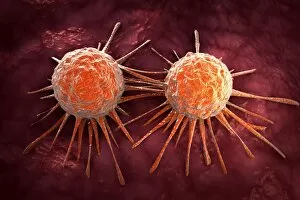
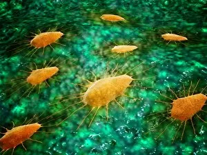
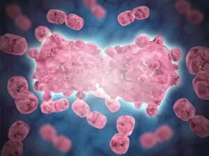
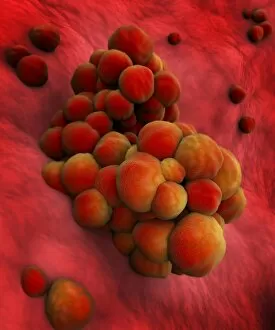
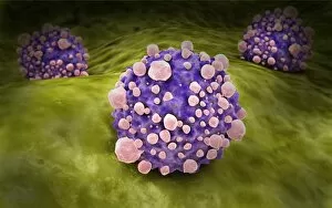
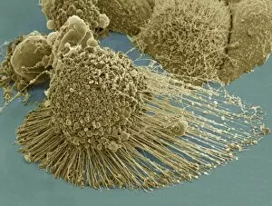
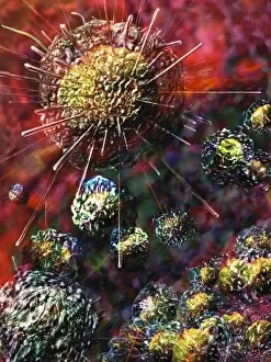
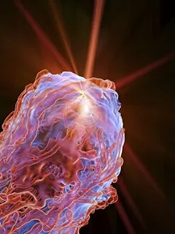
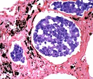
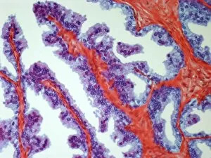
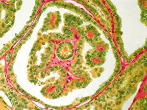
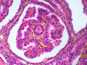
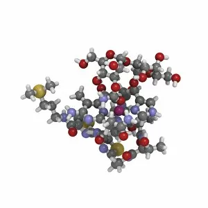
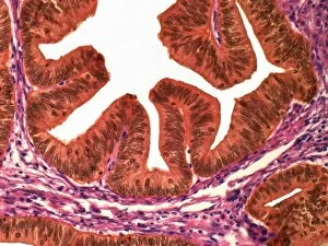
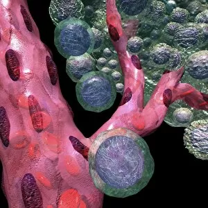
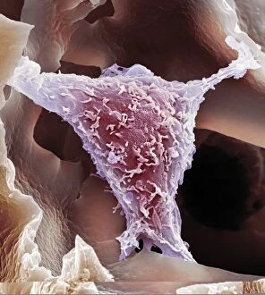
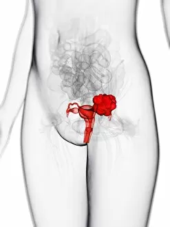
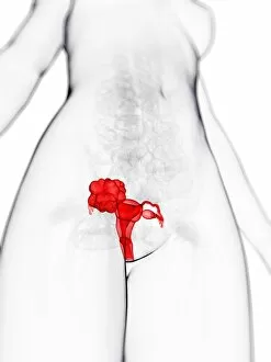
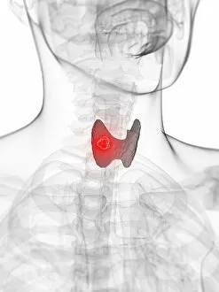
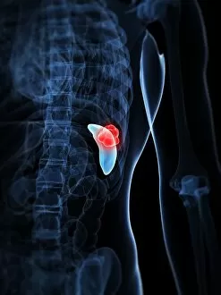
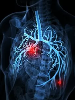
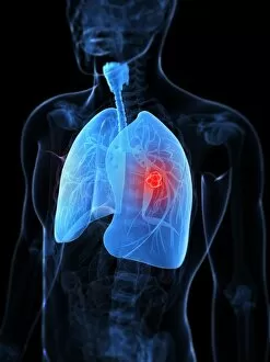
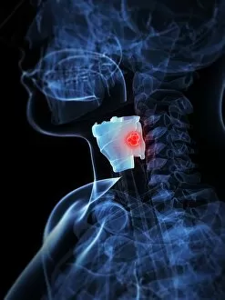
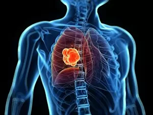
Oncology: Unveiling the Battle Within In the realm of oncology, a fierce war is waged between T lymphocytes and cancer cells. SEM C001 / 1679 captures this epic struggle at a microscopic level, showcasing the intricate dance between these two adversaries. HeLa cells, depicted in light micrographs C017 / 8299 and C017 / 8298, offer us glimpses into their mysterious world. These immortal cells have played an instrumental role in countless scientific breakthroughs, unraveling the secrets of cancer biology. Acute promyelocytic leukemia reveals its sinister face through a haunting micrograph. The image serves as a reminder of the relentless fight against blood cancers that oncologists tirelessly engage in each day. Ovarian cancer's treacherous nature is exposed under the lens of light micrograph C015 / 7103. This glimpse into its cellular landscape fuels our determination to develop effective treatments for this formidable disease. The paclitaxel drug molecule emerges as a potent weapon in our arsenal against cancer. Its molecular structure holds promise for patients battling various forms of malignancies. Within operating rooms worldwide, surgeons skillfully navigate breast cancer cases with precision and expertise. Armamentarii Chirurgici provides us with an intimate view into these life-saving procedures. Cancerous scrofula reminds us that skin diseases can also be harbingers of deeper battles within our bodies. Description des Maladies de la Peau offers insight into dermatological manifestations linked to underlying malignancies. Traite Complet de l'Anatomie de l unveils surgical interventions on malignant tumors – visual proof that medical professionals are at the forefront of combating this pervasive disease. Medical illustrations depicting pilonidal cysts near the natal cleft or stages of colon cancer serve as educational tools to raise awareness about lesser-known aspects while empowering individuals to seek timely medical intervention.