Oncology Collection (#3)

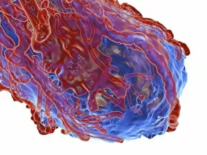
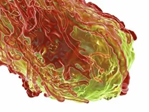
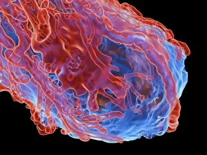
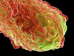
Oncology: Unveiling the Battle Within In the realm of oncology, a fierce war is waged between T lymphocytes and cancer cells

Background imageOncology Collection: Human antitumour antibody molecule

Human antitumour antibody molecule
Human antitumour antibody. Molecular model showing the antitumour antibody BR96 complexed with part of the Lewis antigen. The Lewis antigen is expressed on the surface of human carcinoma cells

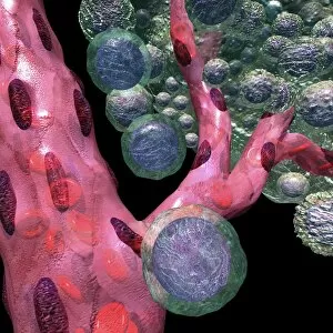
Background imageOncology Collection: Blood vessel formation, artwork C016 / 9842

Blood vessel formation, artwork C016 / 9842
Blood vessel formation. Artwork showing malignant (cancerous) tumour cells (green) promoting the formation of new blood vessels (pink), a process known as angiogenesis
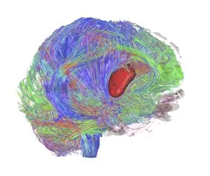
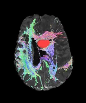
Background imageOncology Collection: Brain tumour, DTI MRI scan C017 / 7057

Brain tumour, DTI MRI scan C017 / 7057
Brain tumour. 3D diffusion tensor imaging (DTI) magnetic resonance imaging (MRI) scan of nerve pathways (coloured) in a brain with a tumour (red, centre right)

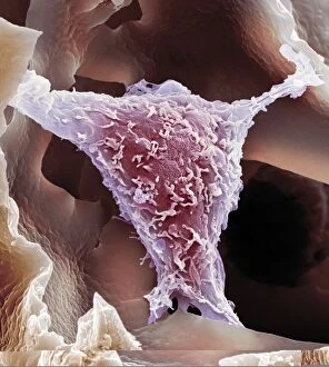
Background imageOncology Collection: HeLa cell, SEM C014 / 0371

HeLa cell, SEM C014 / 0371
HeLa cell. Coloured scanning electron micrograph (SEM) of a HeLa cell (centre) grown in a 3D matrix (background). HeLa cells are a continuously cultured cell line of immortal human cancer cells

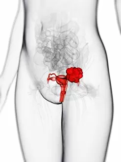
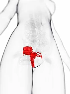
Background imageOncology Collection: Uterine cancer, artwork F006 / 2550

Uterine cancer, artwork F006 / 2550
Uterine cancer, computer artwork

Background imageOncology Collection: Uterine cancer, artwork F006 / 2551

Uterine cancer, artwork F006 / 2551
Uterine cancer, computer artwork

Background imageOncology Collection: Healthy thyroid, artwork F006 / 2526

Healthy thyroid, artwork F006 / 2526
Healthy thyroid, computer artwork

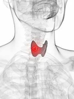
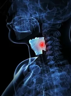
Background imageOncology Collection: Thyroid cancer, artwork F006 / 2527

Thyroid cancer, artwork F006 / 2527
Thyroid cancer, computer artwork

Background imageOncology Collection: Stomach cancer, artwork F006 / 2513

Stomach cancer, artwork F006 / 2513
Stomach cancer, computer artwork

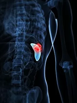
Background imageOncology Collection: Spleen cancer, artwork F006 / 2505

Spleen cancer, artwork F006 / 2505
Spleen cancer, computer artwork

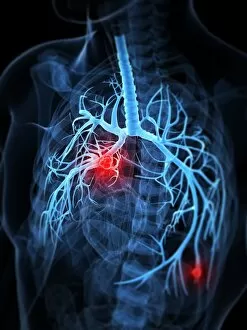
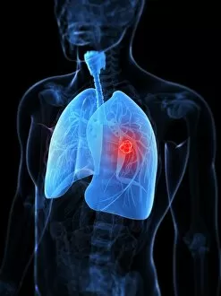
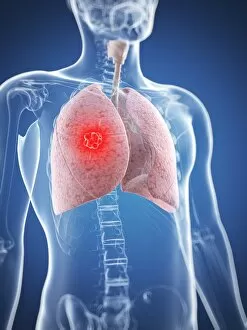
Background imageOncology Collection: Lung cancer, artwork F006 / 2376

Lung cancer, artwork F006 / 2376
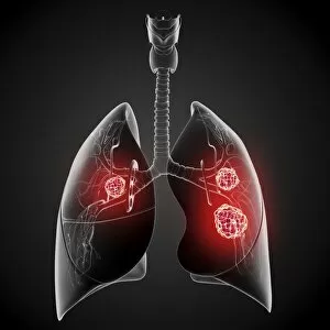
Lung cancer, computer artwork

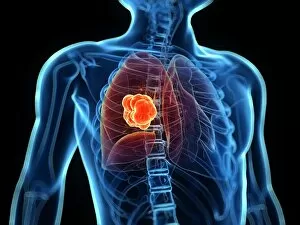
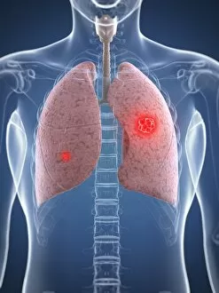
Background imageOncology Collection: Lung cancer, artwork F006 / 2375

Lung cancer, artwork F006 / 2375
Lung cancer, computer artwork

Background imageOncology Collection: Lung cancer, artwork F006 / 2377

Lung cancer, artwork F006 / 2377
Lung cancer, computer artwork

Background imageOncology Collection: Lung cancer, artwork F006 / 2374

Lung cancer, artwork F006 / 2374
Lung cancer, computer artwork

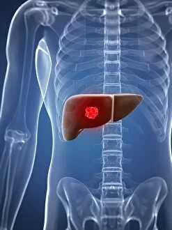
Background imageOncology Collection: Liver cancer, artwork F006 / 2371

Liver cancer, artwork F006 / 2371
Liver cancer, computer artwork

Background imageOncology Collection: Lung cancer, artwork F006 / 2373

Lung cancer, artwork F006 / 2373
Lung cancer, computer artwork

Background imageOncology Collection: Healthy thyroid, artwork

Healthy thyroid, artwork
Healthy thyroid, computer artwork

Background imageOncology Collection: Kidney cancer, artwork F006 / 2357

Kidney cancer, artwork F006 / 2357
Kidney cancer, computer artwork

Background imageOncology Collection: Lung cancer, artwork F006 / 2312

Lung cancer, artwork F006 / 2312
Lung cancer, computer artwork

Background imageOncology Collection: Lung cancer, artwork F006 / 2282

Lung cancer, artwork F006 / 2282
Lung cancer, computer artwork

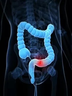
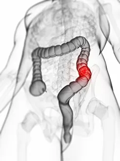
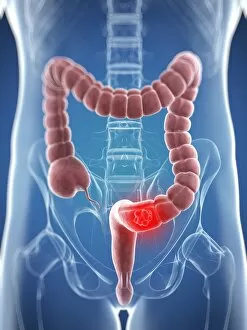
Background imageOncology Collection: Colon cancer, artwork F006 / 2231

Colon cancer, artwork F006 / 2231
Colon cancer, computer artwork

Background imageOncology Collection: Colon cancer, artwork F006 / 2227

Colon cancer, artwork F006 / 2227
Colon cancer, computer artwork

Background imageOncology Collection: Colon cancer, artwork F006 / 2226

Colon cancer, artwork F006 / 2226
Colon cancer, computer artwork

Background imageOncology Collection: Thyroid cancer, artwork F006 / 2217

Thyroid cancer, artwork F006 / 2217
Thyroid cancer, computer artwork

Background imageOncology Collection: Lung cancer, artwork F006 / 2215

Lung cancer, artwork F006 / 2215
Lung cancer, computer artwork

Background imageOncology Collection: Lung cancer, artwork F006 / 2214

Lung cancer, artwork F006 / 2214
Lung cancer, computer artwork

Background imageOncology Collection: Liver cancer, artwork F006 / 2213

Liver cancer, artwork F006 / 2213
Liver cancer, computer artwork

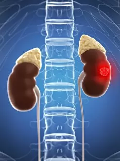
Background imageOncology Collection: Kidney cancer, artwork F006 / 2212

Kidney cancer, artwork F006 / 2212
Kidney cancer, computer artwork

Background imageOncology Collection: Brain cancer, artwork F006 / 2210

Brain cancer, artwork F006 / 2210
Brain cancer, computer artwork

Background imageOncology Collection: Colon cancer, artwork F006 / 2211

Colon cancer, artwork F006 / 2211
Colon cancer, computer artwork

Background imageOncology Collection: Lung cancer, artwork F006 / 2209

Lung cancer, artwork F006 / 2209
Lung cancer, computer artwork

Background imageOncology Collection: Brain tumour, DTI MRI scan C017 / 7059

Brain tumour, DTI MRI scan C017 / 7059
Brain tumour. Axial diffusion tensor imaging (DTI) magnetic resonance imaging (MRI) scan of nerve pathways (coloured) in a brain with a tumour (red, upper centre)

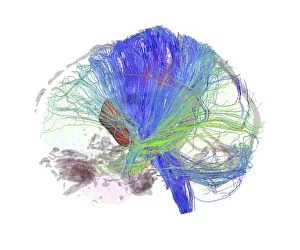
Background imageOncology Collection: Brain tumour, DTI MRI scan C017 / 7058

Brain tumour, DTI MRI scan C017 / 7058
Brain tumour. 3D diffusion tensor imaging (DTI) magnetic resonance imaging (MRI) scan of nerve pathways (coloured) in a brain with a tumour (red, centre left)

Background imageOncology Collection: Tumour, computer artwork F007 / 0218

Tumour, computer artwork F007 / 0218
Tumour, computer artwork. Tumours are caused by the uncontrolled growth of previously normal cells. The resulting growth (green) can invade and damage surrounding tissue

Background imageOncology Collection: Tumour, computer artwork F007 / 0216

Tumour, computer artwork F007 / 0216
Tumour, computer artwork. Tumours are caused by the uncontrolled growth of previously normal cells. The resulting growth (blue) can invade and damage surrounding tissue

Background imageOncology Collection: Tumour, computer artwork F007 / 0214

Tumour, computer artwork F007 / 0214
Tumour, computer artwork. Tumours are caused by the uncontrolled growth of previously normal cells. The resulting growth (green) can invade and damage surrounding tissue

Background imageOncology Collection: Tumour, computer artwork F007 / 0212

Tumour, computer artwork F007 / 0212
Tumour, computer artwork. Tumours are caused by the uncontrolled growth of previously normal cells. The resulting growth (blue) can invade and damage surrounding tissue

Background imageOncology Collection: Bone cancer, light micrograph F006 / 9810

Bone cancer, light micrograph F006 / 9810
Bone cancer. Light micrograph of a section through an undifferentiated pleomorphic sarcoma of bone. This is a malignant (cancerous) tumour. Magnification: x200 when printed at 10 centimetres wide

Background imageOncology Collection: Metastatic breast cancer F006 / 9802

Metastatic breast cancer F006 / 9802
Metastatic breast cancer. Light micrograph of a section through a lymph node showing a malignant (cancerous) tumour (purple) that originated in the breast

Background imageOncology Collection: Female lung cancer, artwork

Female lung cancer, artwork
Female lung cancer, computer artwork

Background imageOncology Collection: Lung cancer, artwork

Lung cancer, artwork
Lung cancer, computer artwork

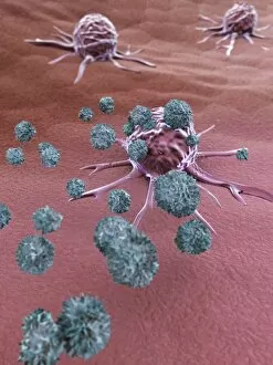
Background imageOncology Collection: NK cells attacking cancer cells, artwork

NK cells attacking cancer cells, artwork
NK cells attacking cancer cells. Computer artwork of human Natural Killer (NK) cells (small, round) attacking cancer cells (large, round)

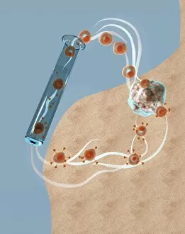
Background imageOncology Collection: Immunotherapy, artwork

Immunotherapy, artwork
Immunotherapy. Computer artwork of immunotherapy being used to destroy a tumour (right). Immunotherapy uses the bodys immune system to fight a disease

Background imageOncology Collection: Cancer cells spreading, artwork

Cancer cells spreading, artwork
Cancer cells spreading. Computer artwork showing cancerous cells (blue) migrating from a tumour (upper centre) into the blood stream (centre)

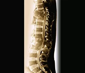
Background imageOncology Collection: Spinal cancer, MRI scan

Spinal cancer, MRI scan
Spinal cancer. Coloured magnetic resonance imaging (MRI) scan of a sagittal section through the cervical (neck) region of the spine of a 22-year-old patient

Background imageOncology Collection: Secondary spinal cancer, MRI scan

Secondary spinal cancer, MRI scan
Secondary spinal cancer. Magnetic resonance imaging (MRI) scan of a section through the spine of a 48-year-old patient, showing malignant (cancerous) tumours

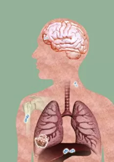
Background imageOncology Collection: Metastatic cancer, artwork

Metastatic cancer, artwork
Metastatic cancer. Computer artwork of a section through a persons upper body and head, showing a primary cancer in the right liver (lower left) that has spread (metastasised)

Background imageOncology Collection: Cancer blood supply, conceptual image C016 / 7505

Cancer blood supply, conceptual image C016 / 7505
Cancer blood supply, conceptual image. Computer artwork representing the blood supply to a cancerous (malignant) tumour


For sale as Licensed Images

Choose your image, Select your licence and Download the media

Oncology: Unveiling the Battle Within In the realm of oncology, a fierce war is waged between T lymphocytes and cancer cells. SEM C001 / 1679 captures this epic struggle at a microscopic level, showcasing the intricate dance between these two adversaries. HeLa cells, depicted in light micrographs C017 / 8299 and C017 / 8298, offer us glimpses into their mysterious world. These immortal cells have played an instrumental role in countless scientific breakthroughs, unraveling the secrets of cancer biology. Acute promyelocytic leukemia reveals its sinister face through a haunting micrograph. The image serves as a reminder of the relentless fight against blood cancers that oncologists tirelessly engage in each day. Ovarian cancer's treacherous nature is exposed under the lens of light micrograph C015 / 7103. This glimpse into its cellular landscape fuels our determination to develop effective treatments for this formidable disease. The paclitaxel drug molecule emerges as a potent weapon in our arsenal against cancer. Its molecular structure holds promise for patients battling various forms of malignancies. Within operating rooms worldwide, surgeons skillfully navigate breast cancer cases with precision and expertise. Armamentarii Chirurgici provides us with an intimate view into these life-saving procedures. Cancerous scrofula reminds us that skin diseases can also be harbingers of deeper battles within our bodies. Description des Maladies de la Peau offers insight into dermatological manifestations linked to underlying malignancies. Traite Complet de l'Anatomie de l unveils surgical interventions on malignant tumors – visual proof that medical professionals are at the forefront of combating this pervasive disease. Medical illustrations depicting pilonidal cysts near the natal cleft or stages of colon cancer serve as educational tools to raise awareness about lesser-known aspects while empowering individuals to seek timely medical intervention.