
Optic Nerve Collection (#2)
"The Optic Nerve: Unveiling the Intricate Pathways of Vision" Step into the fascinating world of the optic nerve


For sale as Licensed Images
Choose your image, Select your licence and Download the media
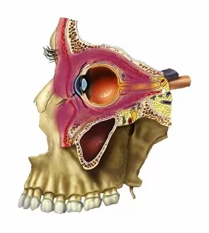
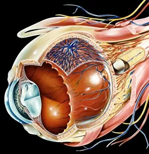
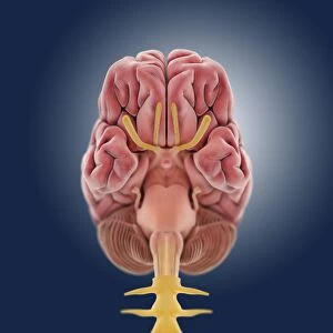
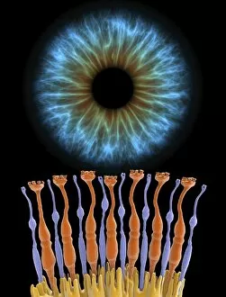
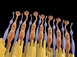
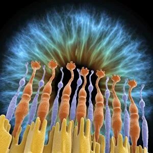
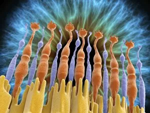
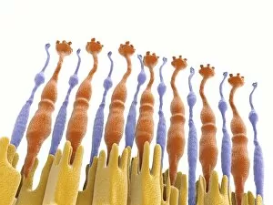
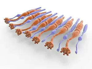
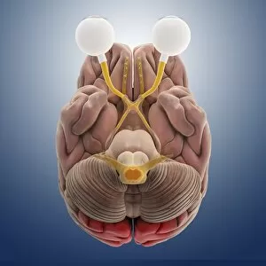
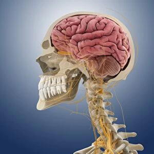
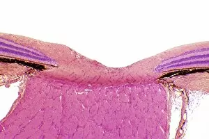
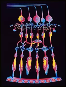
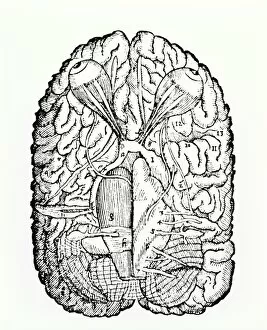
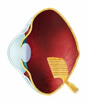
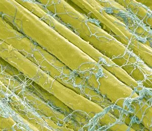
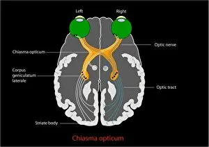
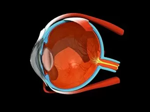
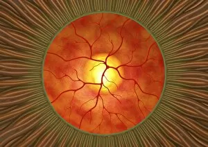
"The Optic Nerve: Unveiling the Intricate Pathways of Vision" Step into the fascinating world of the optic nerve, where intricate networks and delicate structures come together to create our sense of sight. Through a combination of scientific illustrations, artwork, and historical references, we embark on a journey that reveals the wonders hidden within this vital pathway. Retina blood vessels captured under an SEM (Scanning Electron Microscope) showcase their intricate patterns, emphasizing their crucial role in nourishing the retina. Head and neck anatomy artwork provides a broader context for understanding how the optic nerve connects with various regions of our body. Delving deeper into its composition, SEM images reveal optic nerve fibers in all their complexity. These microscopic threads transmit visual information from our eyes to the brain at astonishing speeds. A cross-sectional view unveils normal eye anatomy while highlighting key components such as rods and cones found within the retina. Drawing inspiration from history, we explore Rene Descartes' groundbreaking ideas on vision dating back to 1692. His concept of hydraulic action in nerves laid foundations for understanding how signals travel through this remarkable system connecting eye and brain. Artwork depicting retinal implants showcases cutting-edge technology aimed at restoring vision for those with impaired sight. This glimpse into modern advancements reminds us of humanity's relentless pursuit to unlock new possibilities within our own bodies. A captivating illustration published in 1898 takes us back in time when scientists were just beginning to unravel the mysteries behind human ocular function. The intricacies depicted serve as testament to centuries-long fascination with understanding how we perceive light. Finally, digital illustrations highlight different areas of cortex receiving input from sense organs like eyes – reminding us that vision is not solely confined to one isolated structure but rather an integrated process involving multiple regions within our extraordinary brains. Exploring these diverse perspectives allows us to appreciate both past discoveries and present innovations surrounding the optic nerve.