
Osteoarthritis Collection
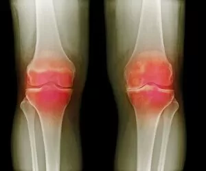
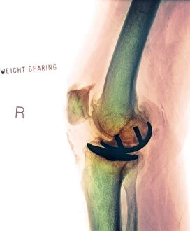
"Osteoarthritis: A Silent Battle Within Our Bones" Arthritic knees, X-ray reveals the hidden pain that osteoarthritis inflicts on our joints


For sale as Licensed Images
Choose your image, Select your licence and Download the media
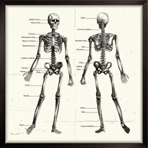
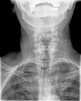
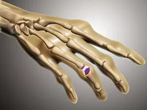
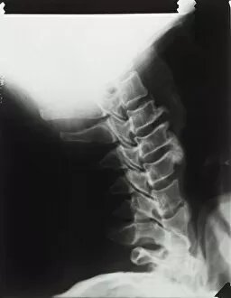
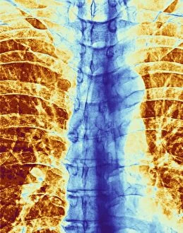
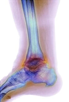
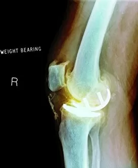
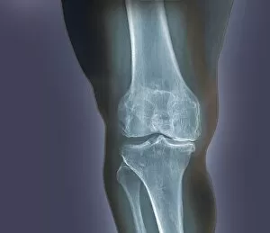
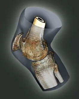
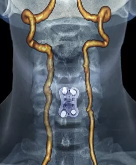
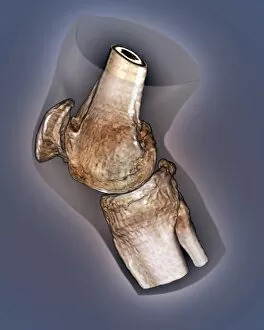
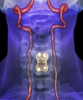
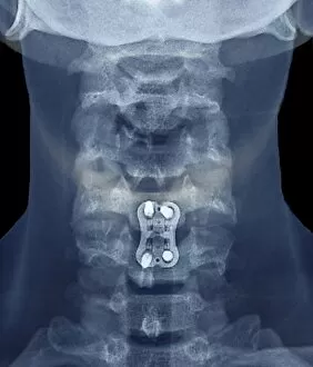
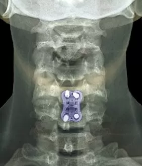
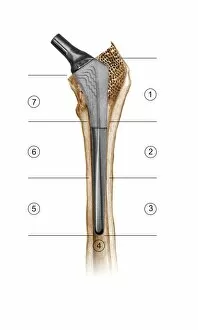
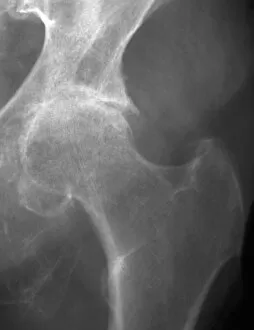
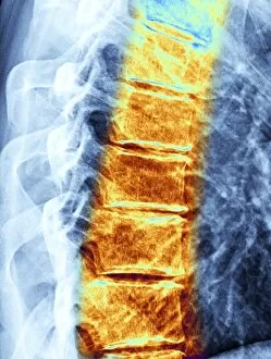
"Osteoarthritis: A Silent Battle Within Our Bones" Arthritic knees, X-ray reveals the hidden pain that osteoarthritis inflicts on our joints. As we age, our once sturdy skeletons become vulnerable to this degenerative disease. A labelled human skeleton engraving serves as a stark reminder of the impact they are have on our musculoskeletal system. The deformation of the cervical spine is evident in an X-ray showcasing arthritis of the neck. In a conceptual image, rheumatoid arthritis manifests its presence in the delicate bones of a human hand. This depiction highlights how different forms of arthritis can affect various parts of our bodies. The thoracic spine bears witness to both scoliosis and osteoarthritis, as seen in X-rays C017/0701 and C017/0704. These images emphasize how these conditions intertwine and contribute to discomfort for those affected. Moving further down the body, X-rays F006/3744 and F006/3745 reveal osteoarthritis taking hold in the hip joint. This common form of arthritis causes pain and stiffness that hinders mobility. Even our ankles are not spared from this debilitating condition; an X-ray labeled F008/3483 shows us firsthand how osteoarthritis affects this crucial joint. Osteoarthritis silently chips away at our quality of life, but with early detection and proper management, we can fight back against its relentless assault on our joints. Let's raise awareness about this chronic condition so that no one has to suffer alone.