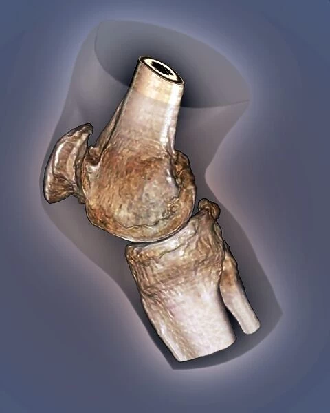
Arthritis of the knee, 3D CT scan C016 / 6511
![]()

Wall Art and Photo Gifts from Science Photo Library
Arthritis of the knee, 3D CT scan C016 / 6511
Arthritis of the knee. Coloured 3D computed tomography (CT) scan of the right knee of a 65 year old patient with severe osteoarthritis. At top is the femur (thigh bone), at bottom is the tibia (shinbone), with the smaller fibula to its right. The patella (knee cap) is at left. Osteoarthritis is a joint disease caused by mechanical wear and tear. In a normal knee, a clear space should be seen between the femur and patellar. Osteoarthritis has led to the loss of the smooth joint surface, and of the separating cartilage. Treatment is with painkillers, or surgery in extreme cases
Science Photo Library features Science and Medical images including photos and illustrations
Media ID 9245391
© ZEPHYR/SCIENCE PHOTO LIBRARY
Arthritic Arthritis Ct Scan Degenerative Femur Joint Knee Osteoarthritis Patella Patient Profile Sagittal Scanner Senior Shinbone Sixties Sixty Five Thigh Bone Three Dimensional Tibia Abnormal Condition Disorder Unhealthy
EDITORS COMMENTS
This print showcases the debilitating effects of arthritis on the knee joint. In vivid detail, a 3D CT scan reveals the right knee of a 65-year-old patient suffering from severe osteoarthritis. The image presents an intricate view, with the femur (thigh bone) positioned at the top and the tibia (shinbone) at the bottom, accompanied by its smaller counterpart, the fibula. The patella (knee cap) can be seen on the left side. Osteoarthritis is a degenerative joint disease primarily caused by mechanical wear and tear over time. In contrast to a healthy knee where there should be ample space between the femur and patellar, this condition has eroded both smooth joint surfaces and separating cartilage. The photograph highlights not only medical aspects but also emphasizes human experiences as it depicts an adult in their sixties grappling with this abnormality that affects their mobility and overall well-being. Treatment options for osteoarthritis range from pain management through medication to surgical intervention in extreme cases. ZEPHYR/SCIENCE PHOTO LIBRARY has skillfully captured this poignant image that serves as a powerful reminder of how arthritis can impact individuals' lives while shedding light on advancements in medical imaging technology like three-dimensional computed tomography scans.
MADE IN THE USA
Safe Shipping with 30 Day Money Back Guarantee
FREE PERSONALISATION*
We are proud to offer a range of customisation features including Personalised Captions, Color Filters and Picture Zoom Tools
SECURE PAYMENTS
We happily accept a wide range of payment options so you can pay for the things you need in the way that is most convenient for you
* Options may vary by product and licensing agreement. Zoomed Pictures can be adjusted in the Cart.

