
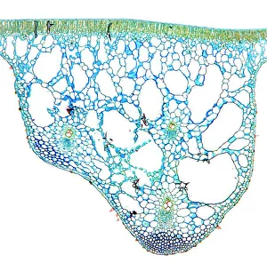
Palisade Mesophyll Collection
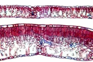
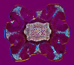
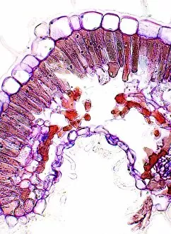
The palisade mesophyll is a fascinating feature found in various plant species, as depicted by the water lily leaf, light micrograph

For sale as Licensed Images
Choose your image, Select your licence and Download the media
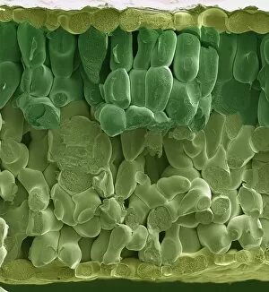
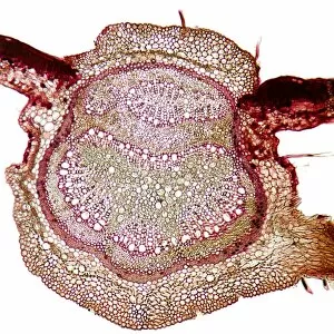
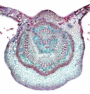
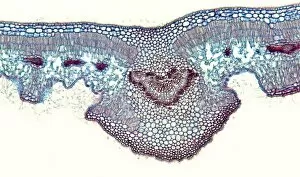
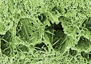
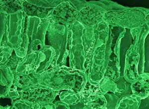
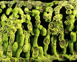
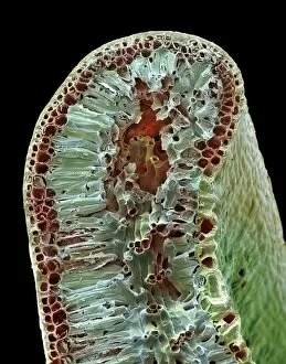
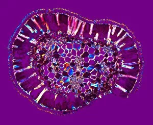
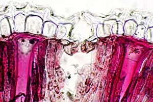
The palisade mesophyll is a fascinating feature found in various plant species, as depicted by the water lily leaf, light micrograph. This specialized tissue plays a crucial role in photosynthesis and can be observed under different microscopic techniques. In the SEM image of a spinach leaf, we can clearly see the densely packed palisade cells arranged vertically to maximize sunlight absorption. Similarly, the beech tree leaf's light micrograph showcases this intricate network of elongated cells that are responsible for capturing solar energy efficiently. Another example is seen in the oleander leaf's light micrograph, where these distinct palisade cells are visible beneath the upper epidermis. The repetitive pattern of tightly packed chloroplast-rich cells indicates their primary function in converting sunlight into chemical energy through photosynthesis. Interestingly, both sycamore and additional beech tree leaves exhibit similar structures when examined under a light microscope. These images reveal rows of closely aligned palisade mesophyll cells nestled between upper and lower epidermal layers. Further exploration using SEM provides more detailed insights into this vital tissue. For instance, an SEM image of a lily leaf demonstrates how individual palisade cells possess numerous chloroplasts within their elongated bodies – an adaptation that enhances their ability to capture sunlight effectively. A closer examination via SEM also allows us to observe cross-sections of leaves with exceptional clarity. In one such image labeled "leaf section, " we witness multiple layers comprising not only palisade mesophyll but also other essential tissues like spongy mesophyll and vascular bundles. Overall, these captivating visuals highlight the diverse forms and functions across various plant species. From water lilies to spinach leaves or even oleander plants – each showcasing unique adaptations for optimal photosynthetic activity – it becomes evident that this specialized tissue plays an indispensable role in sustaining life on our planet through its remarkable ability to harness the power of sunlight.