Pincushion leaf, light micrograph
![]()

Wall Art and Photo Gifts from Science Photo Library
Pincushion leaf, light micrograph
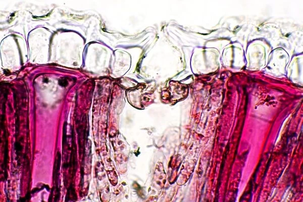
Pin cushion leaf. Polarised light micrograph of a transverse section through a pinchusion (Hakea laurina) leaf. The leaf of this plant has a number of adaptations that help it to minimise water loss through transpiration. It is covered in a thick cuticle (see-through, top) and its stomata (leaf pores, one seen at centre) are sunken with a cup above them that traps moist air. Also seen are palisade mesophyll cells (red), which contain chloroplasts the site of photosynthesis, and radial sclereid cells (bright pink), thick-walled cells that provide structural support. Magnification: x230 when printed at 10 centimetres wide
Science Photo Library features Science and Medical images including photos and illustrations
Media ID 6281414
© DR KEITH WHEELER/SCIENCE PHOTO LIBRARY
Cuticle Palisade Mesophyll Pin Cushion Plant Anatomy Plant Cell Polarised Light Micrograph Stoma Stomata Transverse Section Xerophyte Cells Light Micrograph Light Microscope Sectioned
EDITORS COMMENTS
This print showcases the intricate beauty of a Pincushion leaf, captured under polarised light micrograph. The transverse section through this Hakea laurina leaf reveals its remarkable adaptations that enable it to minimize water loss through transpiration. The leaf's surface is protected by a thick cuticle, providing both transparency and protection. Sunken stomata, visible at the center of the image, are accompanied by cup-like structures that effectively trap moist air. The vibrant red palisade mesophyll cells steal the spotlight in this image as they contain chloroplasts – essential for photosynthesis. These cells play a crucial role in converting sunlight into energy for the plant's survival. Additionally, bright pink radial sclereid cells can be observed throughout the leaf section; these thick-walled cells provide structural support to ensure stability. Printed at 10 centimeters wide with a magnification of x230, this photograph allows us to appreciate nature's microscopic wonders up close. It serves as a reminder of how plants have evolved complex mechanisms to thrive in their environments and adapt to challenging conditions such as limited water availability. This mesmerizing botanical snapshot offers an intriguing glimpse into plant anatomy and biology while highlighting the unique features of this particular species - making it an ideal addition for any nature enthusiast or science lover seeking inspiration from our natural world.
MADE IN THE USA
Safe Shipping with 30 Day Money Back Guarantee
FREE PERSONALISATION*
We are proud to offer a range of customisation features including Personalised Captions, Color Filters and Picture Zoom Tools
SECURE PAYMENTS
We happily accept a wide range of payment options so you can pay for the things you need in the way that is most convenient for you
* Options may vary by product and licensing agreement. Zoomed Pictures can be adjusted in the Cart.

