
Phalanges Collection (#2)
"Unveiling the Marvels of Phalanges: A Journey into the Intricacies of Human Anatomy" Step into the world of phalanges


For sale as Licensed Images
Choose your image, Select your licence and Download the media
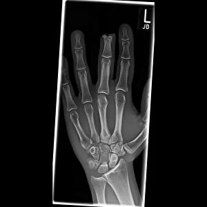
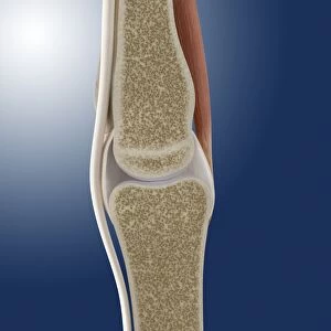
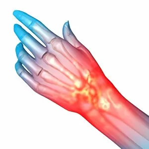
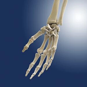
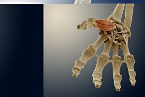
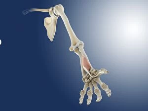
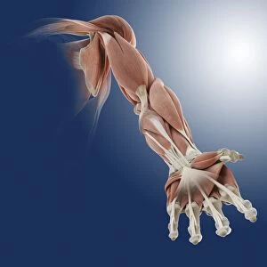
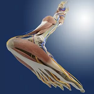
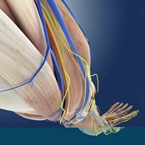
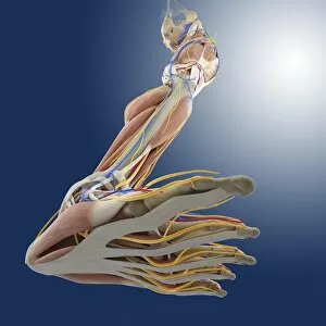
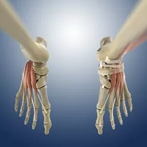
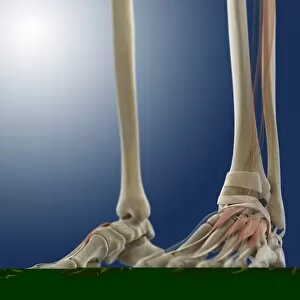
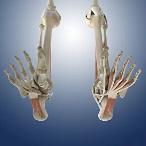
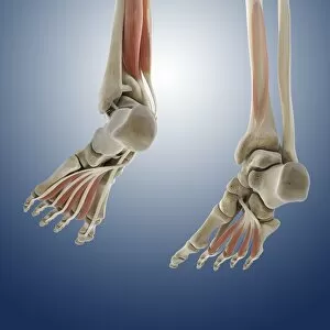
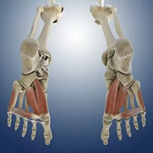
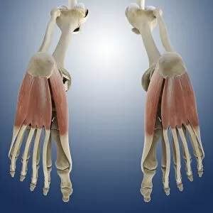
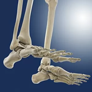
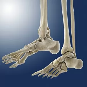
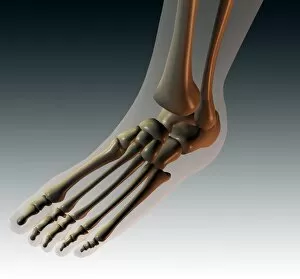
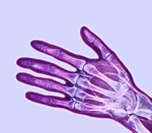
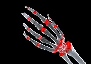
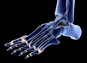
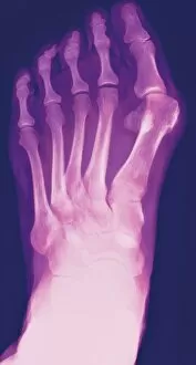
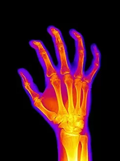
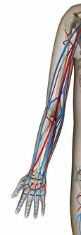
"Unveiling the Marvels of Phalanges: A Journey into the Intricacies of Human Anatomy" Step into the world of phalanges, where we explore the wonders hidden within our normal foot and its intricate skeletal structure. Peering through an X-ray lens, witness the mesmerizing beauty of a skeleton from below, revealing the delicate balance between bones and joints. Delve deeper with detailed diagrams showcasing the complex network of bones in our hands and arms, unraveling their vital role in everyday activities. Behold a coloured X-ray masterpiece capturing the essence of a healthy man's hand – a true testament to nature's flawless design. Embark on an anatomical adventure as we unveil diagrams depicting the bones that support our right leg and hip, enabling us to stand tall. Explore captivating X-rays that showcase healthy human hands in all their glory – marvel at their intricate composition and functionality. Travel back in time with an Australopithecine or Homo habilis foot cast (OH8), offering insights into our evolutionary journey towards bipedalism. Admire stunning artwork illustrating outer ankle ligaments (C013 / 4452) – discover how these crucial structures provide stability during movement. Immerse yourself further with artwork showcasing inner ankle ligaments (C013 / 4451), shedding light on another aspect of this remarkable joint's anatomy. Witness innocence through a child hand X-ray - see how tiny fingers hold immense potential for growth and exploration. Take a historical leap as we present a vibrant lithograph comparing hand bones across nine different mammals from 1898 - highlighting both similarities and unique adaptations. Thumb your way through life’s intricacies with a colourful lithograph dedicated solely to this essential digit - celebrating its versatility like never before.