Physiology Collection (#2)

"Unlocking the Secrets of the Human Body

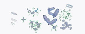
Background imagePhysiology Collection: Illustration of response to infection after vaccination, involving microbe, antigens, antibody, plas

Illustration of response to infection after vaccination, involving microbe, antigens, antibody, plas

Background imagePhysiology Collection: Cross section biomedical illustration of iliac crest site on pelvis

Cross section biomedical illustration of iliac crest site on pelvis

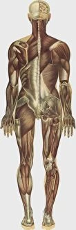
Background imagePhysiology Collection: The human body with superimposed colored plates by Julien Bougle

The human body with superimposed colored plates by Julien Bougle
The human body with superimposed colored plates, by Julien Bougle, circa 1899

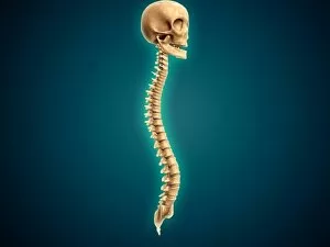
Background imagePhysiology Collection: Conceptual image of human skull and spinal cord

Conceptual image of human skull and spinal cord

Background imagePhysiology Collection: Human fetus inside amniotic sac

Human fetus inside amniotic sac

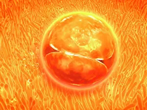
Background imagePhysiology Collection: Embryo development 24-36 hours after fertilization

Embryo development 24-36 hours after fertilization

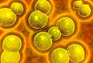
Background imagePhysiology Collection: Microscopic view of chlamydia

Microscopic view of chlamydia. Chlamydia is a common sexually transmitted disease (STD) caused by a bacterium

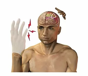
Background imagePhysiology Collection: Phantom pain after amputation, artwork

Phantom pain after amputation, artwork
Phantom pain after amputation. Artwork of a man experiencing phantom pain (red flash) from his amputated hand (left). This is caused by stimuli to the motor and pre-motor cortical areas of his brain

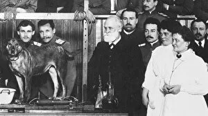
Background imagePhysiology Collection: (1849-1936). Russian physiologist. Pavlov (center, with beard) with assistants

(1849-1936). Russian physiologist. Pavlov (center, with beard) with assistants
IVAN PETROVICH PAVLOV (1849-1936). Russian physiologist. Pavlov (center, with beard) with assistants and students at the Imperial Military Academy of Medicine, St

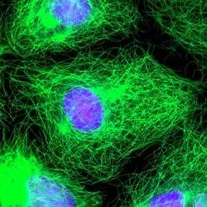
Background imagePhysiology Collection: Cell division, fluorescent micrograph

Cell division, fluorescent micrograph
Cell division. Immunofluorescent light micrograph of a human epithelial cell (centre) during the interphase stage of mitosis

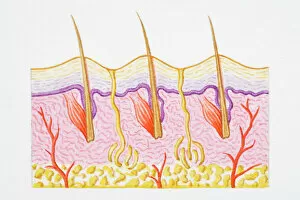
Background imagePhysiology Collection: Illustration of cross section of human skin with heat trapped by erect hairs

Illustration of cross section of human skin with heat trapped by erect hairs

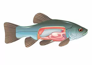
Background imagePhysiology Collection: Digital cross section illustration of fish showing gas bladder which contributes to the ability to

Digital cross section illustration of fish showing gas bladder which contributes to the ability to control buoyancy

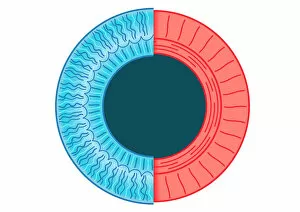
Background imagePhysiology Collection: Digital illustration of dilated pupil of human eye with contraction of outer radial muscle fibres

Digital illustration of dilated pupil of human eye with contraction of outer radial muscle fibres

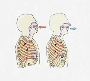
Background imagePhysiology Collection: Illustration showing diaphragm moving down when exhaling and up when inhaling

Illustration showing diaphragm moving down when exhaling and up when inhaling

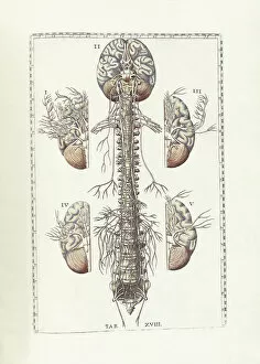
Background imagePhysiology Collection: The science of human anatomy by Bartholomeo Eustachi

The science of human anatomy by Bartholomeo Eustachi, depicting the shape, size and relative position of the organs of the human body. Circa 18th century

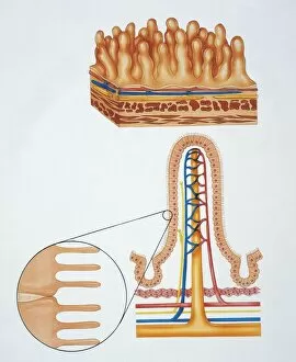
Background imagePhysiology Collection: Illustration showing intestinal villi

Illustration showing intestinal villi
Human anatomy: intestinal villi. Drawing

Background imagePhysiology Collection: Digital cross section illustration of human cerebellar cortex

Digital cross section illustration of human cerebellar cortex

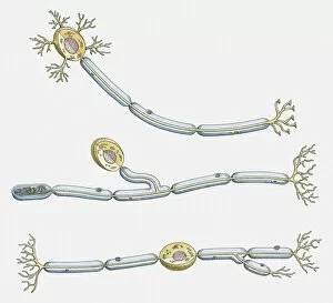
Background imagePhysiology Collection: Illustration of multipolar, unipolar, and bipolar neuron

Illustration of multipolar, unipolar, and bipolar neuron

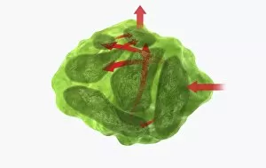
Background imagePhysiology Collection: Digital illustration of direction of information in amygdala

Digital illustration of direction of information in amygdala

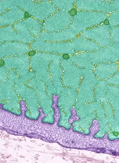
Background imagePhysiology Collection: Eye muscle, TEM C014 / 1468

Eye muscle, TEM C014 / 1468
Eye muscle. Transmission electron micrograph (TEM) of a section through a striated muscle cell from the ciliary muscle of a human eye

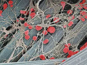
Background imagePhysiology Collection: Blood clot on plaster, SEM

Blood clot on plaster, SEM
Blood clot on plaster. Coloured scanning electron micrograph (SEM) of blood clotting on the surface of a sticking plaster used to dress a small cut

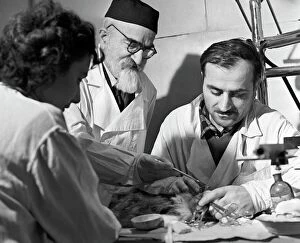
Background imagePhysiology Collection: Beritashvili and students, Tbilisi, 1962

Beritashvili and students, Tbilisi, 1962
Ivan Beritashvili (1885-1974, centre), Soviet physiologist, instructing students who are studying the brain of a laboratory animal

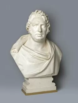
Background imagePhysiology Collection: Bust of John Fuller, philanthropist

Bust of John Fuller, philanthropist
Marble bust of John Fuller (1757-1834), English philanthropist. Fuller was born at North Stoneham, Hampshire. At the age of four he lost his father, the local clergyman
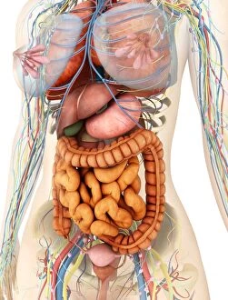
Background imagePhysiology Collection: Female body showing digestive and circulatory system

Female body showing digestive and circulatory system
Medical illustration of female body showing digestive and circulatory system, perspective view

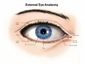
Background imagePhysiology Collection: External anatomy of the human eye (with labels)

External anatomy of the human eye (with labels)

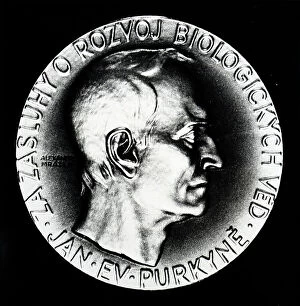
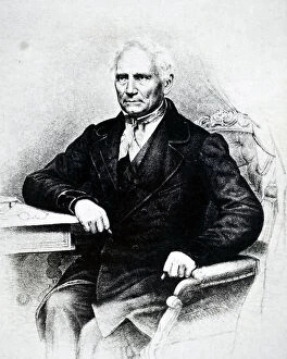
Background imagePhysiology Collection: Jan Evangelista Purkyne, 19th century (engraving)

Jan Evangelista Purkyne, 19th century (engraving)
3733542 Jan Evangelista Purkyne, 19th century (engraving); (add.info.: Portrait of Jan Evangelista Purkyne (1787-1869) a Czech anatomist and physiologist

Background imagePhysiology Collection: Lecture on physiology and hygiene in progress at the Young Men's Christian Association (YMCA's)

Lecture on physiology and hygiene in progress at the Young Men's Christian Association (YMCA's) Exeter Hall
530224 Lecture on physiology and hygiene in progress at the Young Men's Christian Association (YMCA's) Exeter Hall, London. Wood engraving 1887; Universal History Archive/UIG

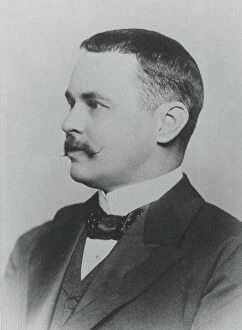
Background imagePhysiology Collection: Ronald Ross (1857-1932) British physician, c.1902 (b/w photo)

Ronald Ross (1857-1932) British physician, c.1902 (b/w photo)
527308 Ronald Ross (1857-1932) British physician, c.1902 (b/w photo); (add.info.: Discovered the Malaria parasite and its life cycle)

Background imagePhysiology Collection: Stephen Hales experiment to demonstrate circulation in plants

Stephen Hales experiment to demonstrate circulation in plants
5310239 Stephen Hales experiment to demonstrate circulation in plants; (add.info.: Engraving depicting Stephen Hales experiment to demonstrate circulation in plants)

Background imagePhysiology Collection: The top layer of muscles of a male body

The top layer of muscles of a male body
5307956 The top layer of muscles of a male body.; (add.info.: Illustration depicting a front and rear view of the top layer of muscles of a male body)

Background imagePhysiology Collection: Santorio Santorio

Santorio Santorio
5307546 Santorio Santorio.; (add.info.: Engraving depicting Santorio Santorio seated in his balance (a steelyard) in which he could eat and sleep

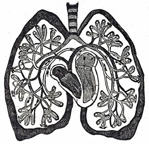
Background imagePhysiology Collection: Diagram of the circulation of blood as understood after William Harvey's work

Diagram of the circulation of blood as understood after William Harvey's work
5309470 Diagram of the circulation of blood as understood after William Harvey's work, showing the Lesser Circulation (Pulmonary Circulation)

Background imagePhysiology Collection: Jan Evangelista Purkyne, 19th century (engraving)

Jan Evangelista Purkyne, 19th century (engraving)
3733543 Jan Evangelista Purkyne, 19th century (engraving); (add.info.: Portrait of Jan Evangelista Purkyne (1787-1869) a Czech anatomist and physiologist

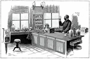
Background imagePhysiology Collection: Robert Koch (1843-1910) German bacteriologist and physician in his laboratory

Robert Koch (1843-1910) German bacteriologist and physician in his laboratory
530414 Robert Koch (1843-1910) German bacteriologist and physician in his laboratory. In 1890 Koch introduced Tuberculin which he thought was a cure for Tuberculosis

Background imagePhysiology Collection: Title page of Haller's Deux memoires sur le mouvement du sang, 1756

Title page of Haller's Deux memoires sur le mouvement du sang, 1756
6317520 Title page of Haller's Deux memoires sur le mouvement du sang, 1756; (add.info.: Title page of Haller's Deux memoires sur le mouvement du sang, 1756)

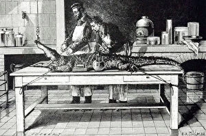
Background imagePhysiology Collection: An engraving depicting Paul Bert determining the biting power of a crocodile - Shamone

An engraving depicting Paul Bert determining the biting power of a crocodile - Shamone
5311545 An engraving depicting Paul Bert determining the biting power of a crocodile - Shamone. The animals lower jaw was fixed and a rope tied the upper one to a dynamometer
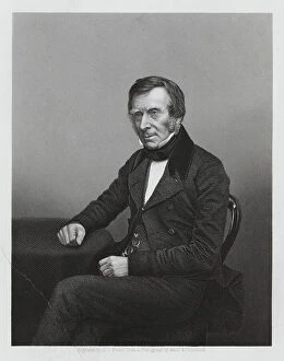
Background imagePhysiology Collection: Sir Benjamin Collins Brodie, 1st Baronet, English physiologist and surgeon (engraving)

Sir Benjamin Collins Brodie, 1st Baronet, English physiologist and surgeon (engraving)
8677064 Sir Benjamin Collins Brodie, 1st Baronet, English physiologist and surgeon (engraving) by Pound, Daniel John. (fl.1860); Private Collection; (add.info.: Sir Benjamin Collins Brodie)
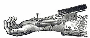
Background imagePhysiology Collection: An engraving depicting a sphygmograph, 19th century (engraving)

An engraving depicting a sphygmograph, 19th century (engraving)
5311400 An engraving depicting a sphygmograph, 19th century (engraving); (add.info.: a device invented by Etienne-Jules Marey for recording the pulse and variations graphically in blood and pressure)

Background imagePhysiology Collection: Kneeling Academic Male Nude, n.d. Creator: Antoine Dumont

Kneeling Academic Male Nude, n.d. Creator: Antoine Dumont
Kneeling Academic Male Nude, n.d

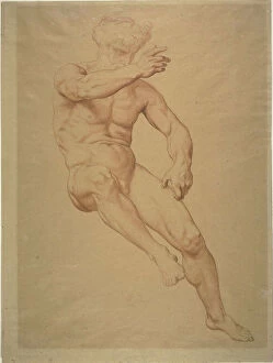
Background imagePhysiology Collection: Academic Nude, n.d. Creator: Alexandre Cabanel

Academic Nude, n.d. Creator: Alexandre Cabanel
Academic Nude, n.d

Background imagePhysiology Collection: Professor Franciscus Donders (1818-1889), Physiologist and Opthalmologist, 1888

Professor Franciscus Donders (1818-1889), Physiologist and Opthalmologist, 1888

Background imagePhysiology Collection: Class in physiology, 1904. Creator: Frances Benjamin Johnston

Class in physiology, 1904. Creator: Frances Benjamin Johnston
Class in physiology, 1904

Background imagePhysiology Collection: Physiologie du Buveur: ls quatre ages, 1862. Creator: Charles Maurand

Physiologie du Buveur: ls quatre ages, 1862. Creator: Charles Maurand
Physiologie du Buveur: ls quatre ages, 1862

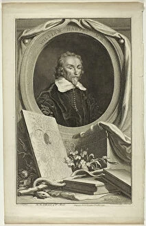
Background imagePhysiology Collection: William Harvey, M.D. 1739. Creator: Jacobus Houbraken

William Harvey, M.D. 1739. Creator: Jacobus Houbraken
William Harvey, M.D. 1739

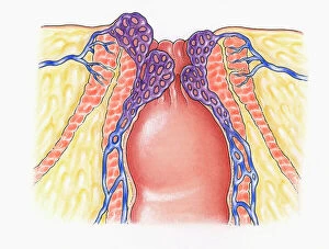
Background imagePhysiology Collection: Cross section illustration of human anal column showing external and internal sphincters

Cross section illustration of human anal column showing external and internal sphincters, healthy veins
Anal, Anatomy, Anus, Biomedical Illustration, Blood Flow, Close-Up, Column, Cross Section, External, Haemorrhoids, Healthcare and Medicine, Healthy, Human Vein, Inflammation, Internal, No People

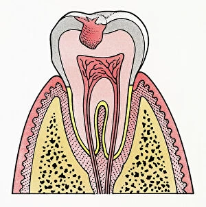
Background imagePhysiology Collection: Cross section illustration showing tooth decay

Cross section illustration showing tooth decay
Anatomy, Bacterium, Biomedical Illustration, Blood Supply, Close-Up, Cross Section, Decay, Dental Hygiene, Enamel, Full Length, Healthcare and Medicine, Human Bone, Human Jaw Bone, Human Teeth

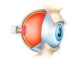
Background imagePhysiology Collection: Cross section illustration of anatomy of human eye showing use of optic nerve

Cross section illustration of anatomy of human eye showing use of optic nerve
Anatomy, Biomedical Illustration, Close-Up, Cross Section, Eyelash, Fovea, Human Eye, Iris, No People, Optic Nerve, Part Of, Physiology, Pupil, Sensory Perception, Side View, Single Object


For sale as Licensed Images

Choose your image, Select your licence and Download the media

"Unlocking the Secrets of the Human Body: Exploring Physiology from Head to Toe" Dive into the intricate world of human physiology with a captivating view of the anatomy of the brain, seen from an inferior perspective. Step back in time and witness Joseph Wright's masterpiece, "The Airpump, " as it symbolizes our quest to understand how our bodies function on a physiological level. Discover the hidden powerhouses behind every smile and frown with a detailed diagram showcasing facial muscles labeled for your convenience. Take a front-row seat to unravel the complexity of facial muscles as they work harmoniously together, providing us with countless expressions that define our humanity. Delve into knee joint mechanics and gain insight into how this remarkable structure allows us to walk, run, and perform various movements effortlessly. Journey beneath your skin's surface and explore the fascinating relationship between hair follicles and skin – two interconnected systems that contribute to our unique appearance. Witness synapse nerve junctions through a TEM image, offering a glimpse into one of nature's most efficient communication networks within our nervous system. Marvel at the intricacies of head muscles as they enable us to express emotions, chew food, speak articulately, or simply turn our heads towards something captivating. Uncover ancient evolutionary structures within our brains by examining cross-section illustrations depicting limbic system functions alongside primitive forebrain regions. Embark on an enlightening tour through your body's drainage system - discover how lymphatic vessels play an essential role in maintaining overall health and immunity. Explore superior views of colored lobes accompanied by labels; see how different brain regions contribute uniquely to cognition, emotion regulation & sensory processing Dissecting human brain anatomy from lateral view reveals its awe-inspiring complexity; marvel at its convoluted folds & diverse functional areas shaping who we are.