Physiology Collection (#6)

"Unlocking the Secrets of the Human Body

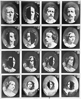
Background imagePhysiology Collection: Physiognomical studies, from 22 to 28, from Mecanisme de la Physionomie humaine ou

Physiognomical studies, from 22 to 28, from Mecanisme de la Physionomie humaine ou
CHT207798 Physiognomical studies, from 22 to 28, from Mecanisme de la Physionomie humaine ou analyse electrophysiologique des passions by Duchenne de Boulogne, published 1876

Background imagePhysiology Collection: Portrait of Etienne Jules Marey (b / w photo)

Portrait of Etienne Jules Marey (b / w photo)
CHT274950 Portrait of Etienne Jules Marey (b/w photo) by Nadar, Paul (1856-1939); Academie des Sciences, Paris, France; (add.info)

Background imagePhysiology Collection: Portrait of Francois Xavier Bichat (1771-1802)

Portrait of Francois Xavier Bichat (1771-1802)
XIR169070 Portrait of Francois Xavier Bichat (1771-1802) by Delafontaine, Pierre Maximilien (1774-1860); 100x81 cm; ChAteau de Versailles, France; French, out of copyright

Background imagePhysiology Collection: Liverpool University George Holt medal for Physiology, 1897 (metal)

Liverpool University George Holt medal for Physiology, 1897 (metal)
1203534 Liverpool University George Holt medal for Physiology, 1897 (metal) by Frampton, George James (1860-1928); diam: 8.9 cm; Leeds Museums and Galleries (Leeds Art Gallery)

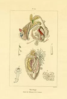
Background imagePhysiology Collection: Intestines, 1833-39 (coloured engraving)

Intestines, 1833-39 (coloured engraving)
999547 Intestines, 1833-39 (coloured engraving) by French School, (19th century); Private Collection; (add.info.: A Dictionnaire pittoresque d histoire naturelle et des phenomAnes de la natureA)

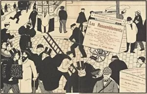
Background imagePhysiology Collection: Book jacket for 'Les Rassemblements', 1896. Creator: Felix Vallotton

Book jacket for "Les Rassemblements", 1896. Creator: Felix Vallotton
Book jacket for " Les Rassemblements", 1896. [Physiologies de la rue par M.M. Paul Adam, Alfred Athys, Victor Barrucand, Tristan Bernard, Leon Blum, Romain Coolus, Felix Feneon

Background imagePhysiology Collection: T A SMITH, SCIENTIST

T A SMITH, SCIENTIST
T A SMITH lecturer on anatomy, physiology and chemistry, but not listed in any reference book : a fine example of a scientist demonstrating Date: flourished 1858

Background imagePhysiology Collection: Fernel, (1497-1558), 1830. Creator: Unknown

Fernel, (1497-1558), 1830. Creator: Unknown
Fernel, (1497-1558), 1830. Jean Francois Fernel (1497-1558) French physician who introduced the term physiology to medicine, was the first person to describe the spinal canal

Background imagePhysiology Collection: W. Harvey, (1578-1657), 1830. Creator: Unknown

W. Harvey, (1578-1657), 1830. Creator: Unknown
W. Harvey, (1578-1657), 1830. William Harvey (1578-1657) English physician educated at Gonville and Caius College, Cambridge

Background imagePhysiology Collection: Galen, (129 AD-c216), 1830. Creator: Unknown

Galen, (129 AD-c216), 1830. Creator: Unknown
Galen, (129 AD-c216), 1830. Aelius Galenus or Claudius Galenus (129 AD-c216) known as Galen of Pergamon, Greek physician, surgeon and philosopher in the Roman Empire

Background imagePhysiology Collection: Leonardo da Vinci (XXXL)

Leonardo da Vinci (XXXL)
Leonardo da Vinci

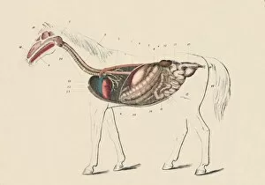
Background imagePhysiology Collection: Longitudinal Section of the Horse, c1879. Creator: Unknown

Longitudinal Section of the Horse, c1879. Creator: Unknown
Longitudinal Section of the Horse, c1879. From " The Book of the Horse", by S. Sidney. [Cassell & Company, Limited, London, Paris, New York, Melbourne]

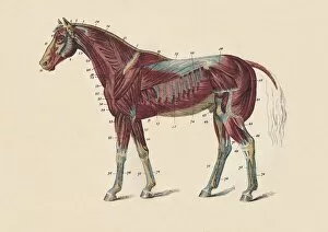
Background imagePhysiology Collection: External Muscles and Tendons of the Horses Body, c1879. Creator: Unknown

External Muscles and Tendons of the Horses Body, c1879. Creator: Unknown
External Muscles and Tendons of the Horses Body, c1879. From " The Book of the Horse", by S. Sidney. [Cassell & Company, Limited, London, Paris, New York, Melbourne]

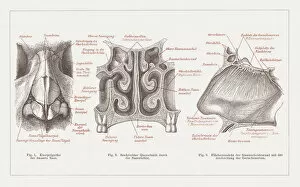
Background imagePhysiology Collection: Anatomy of the human nose, lithograph, published in 1877

Anatomy of the human nose, lithograph, published in 1877
Anatomy of the human nose. Lithograph, published in 1877
Background imagePhysiology Collection: Human skeleton and muscles, hand-coloured engraving, published in 1861

Human skeleton and muscles, hand-coloured engraving, published in 1861
Human skeleton and muscles. Hand-coloured engraving, published in 1861
Background imagePhysiology Collection: Metamorphosis, 19 century science illustration

Metamorphosis, 19 century science illustration
Metamorphosis of insects. A photo of an original hand-colored engraving from Johann Christiaan Seppas Beschouwing der wonderen Gods, in de minst geachte schepzelena published in 1762

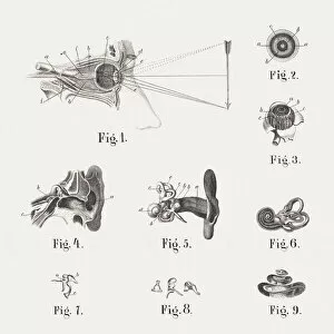
Background imagePhysiology Collection: Anatomy of the human eye and ear, published in 1861

Anatomy of the human eye and ear, published in 1861
Anatomy of the human eye and ear: 1) eye and eye socket, 2) eyeball (cross section, 3) eyeball with open sclera, 4) ear canals (cross section), 5) insulated inner hearing organs with earlobe

Background imagePhysiology Collection: Professor EP Cathcart, CBE. FRS. (Glasgow University), 1926. Artist: Norah Neilson Gray

Professor EP Cathcart, CBE. FRS. (Glasgow University), 1926. Artist: Norah Neilson Gray
Professor EP Cathcart, CBE. FRS. (Glasgow University), 1926. Edward Provan Cathcart (1877-1954), Scottish physician and physiologist. From The Studio Volume 91

Background imagePhysiology Collection: Sir Benjamin Brodie, English doctor, c1860 (1878)

Sir Benjamin Brodie, English doctor, c1860 (1878). Benjamin Collins Brodie (1783-1862) was a physiologist and surgeon who pioneered research into bone and joint disease

Background imagePhysiology Collection: Professor Rene Leriche, French surgeon and physiologist, 1939

Professor Rene Leriche, French surgeon and physiologist, 1939. A photograph from Album de Photographies, Dans L Intimite de Personnages Illustres, 1855-1915, Editions MD, 22 Rue de L Arcade, Paris 8

Background imagePhysiology Collection: Benjamin Ward Richardson, British physician and writer on medical history, 1883

Benjamin Ward Richardson, British physician and writer on medical history, 1883. Artist: Lock & Whitfield
Benjamin Ward Richardson, British physician and writer on medical history, 1883. Richardson was an eminent physician, anaesthetist and physiologist

Background imagePhysiology Collection: William Benjamin Carpenter, CB, MD, LLD, FRS, FLS, FGS, English physiologist and naturalist, 1883

William Benjamin Carpenter, CB, MD, LLD, FRS, FLS, FGS, English physiologist and naturalist, 1883. From Men of Mark: a gallery of contemporary portraits of men distinguished in the Senate

Background imagePhysiology Collection: The Muscular System of the Human Body

The Muscular System of the Human Body - from the front and the rear. Date: circa 1790

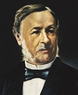
Background imagePhysiology Collection: SCHWANN, Theodor (1810 - 1882). German physiologist

SCHWANN, Theodor (1810 - 1882). German physiologist, anatomist and naturalist. He developed the cell theory. Oil

Background imagePhysiology Collection: Arm flexing bicep with arm bones and bicep revealed

Arm flexing bicep with arm bones and bicep revealed

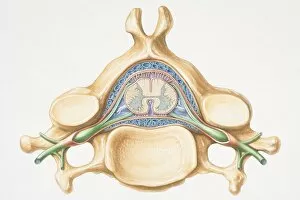
Background imagePhysiology Collection: Anterior cross section of spinal column revealing how vertebra fits round spinal cord

Anterior cross section of spinal column revealing how vertebra fits round spinal cord

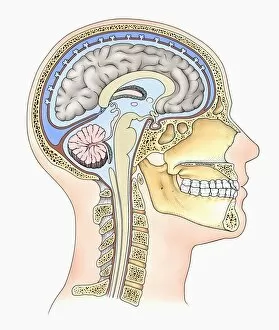
Background imagePhysiology Collection: Lateral cross section through the skull showing the brain with intermediate layers (meninges)

Lateral cross section through the skull showing the brain with intermediate layers (meninges) highlighted

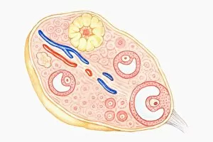
Background imagePhysiology Collection: Cross section illustration of human ovary

Cross section illustration of human ovary

Background imagePhysiology Collection: Illustration of reptile, bird, rabbit and human embryos in early stage of development

Illustration of reptile, bird, rabbit and human embryos in early stage of development

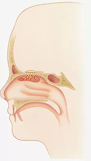
Background imagePhysiology Collection: Illustration of smell sensors, nasal epithelium, olfactory bulb, turbinate, bones

Illustration of smell sensors, nasal epithelium, olfactory bulb, turbinate, bones, frontal sinus and sphenoid sinus in nasal cavity

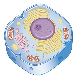
Background imagePhysiology Collection: Illustration of cell nucleus containing cell cytoplasm, mitochondria units, DNA, and chromosome

Illustration of cell nucleus containing cell cytoplasm, mitochondria units, DNA, and chromosome

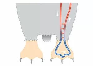
Background imagePhysiology Collection: Digital illustration of penguin circulation showing countercurrent mechanism of warm blood flowing f

Digital illustration of penguin circulation showing countercurrent mechanism of warm blood flowing f

Background imagePhysiology Collection: Cross section digital illustration of Sea Urchin showing gonads, intestine, ampullae and radial cana

Cross section digital illustration of Sea Urchin showing gonads, intestine, ampullae and radial cana

Background imagePhysiology Collection: Digital cross section illustration of male reproductive system

Digital cross section illustration of male reproductive system

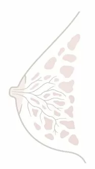
Background imagePhysiology Collection: Digital illustration of human breast anatomy, cross section

Digital illustration of human breast anatomy, cross section

Background imagePhysiology Collection: Digital illustration of showing front view of human brain

Digital illustration of showing front view of human brain

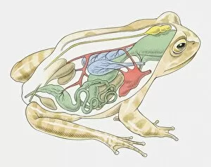
Background imagePhysiology Collection: Illustration of anatomy of frog

Illustration of anatomy of frog

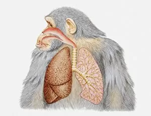
Background imagePhysiology Collection: Illustration of lungs and respiratory system of a chimpanzee

Illustration of lungs and respiratory system of a chimpanzee

Background imagePhysiology Collection: Illustration of female human body showing skeleton, brain, heart, lungs, digestive system

Illustration of female human body showing skeleton, brain, heart, lungs, digestive system, and uterus

Background imagePhysiology Collection: Cross section biomedical illustration of link between the ear, nose and throat

Cross section biomedical illustration of link between the ear, nose and throat

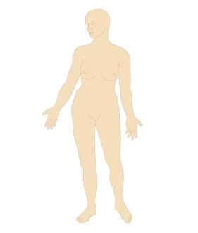
Background imagePhysiology Collection: Digital illustration of body shape of naked adult woman, front view

Digital illustration of body shape of naked adult woman, front view

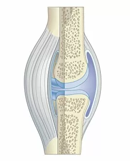
Background imagePhysiology Collection: Cross section biomedical illustration of synovial joint

Cross section biomedical illustration of synovial joint

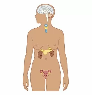
Background imagePhysiology Collection: Cross section biomedical illustration of endocrine system in adult female

Cross section biomedical illustration of endocrine system in adult female

Background imagePhysiology Collection: Medical ilustration of a hiatal hernia in the upper part of the stomach into the thorax

Medical ilustration of a hiatal hernia in the upper part of the stomach into the thorax

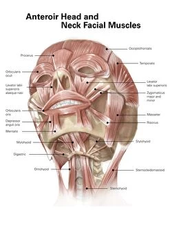
Background imagePhysiology Collection: Anterior neck and facial muscles of the human head (with labels)

Anterior neck and facial muscles of the human head (with labels)

Background imagePhysiology Collection: Anatomy of human respiratory system

Anatomy of human respiratory system

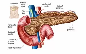
Background imagePhysiology Collection: Anatomy of pancreas

Anatomy of pancreas

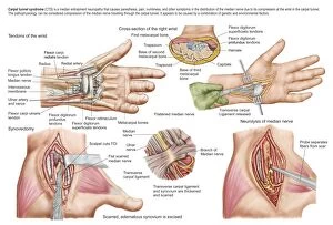
Background imagePhysiology Collection: Medical illustration showing carpal tunnel syndrome in the human wrist

Medical illustration showing carpal tunnel syndrome in the human wrist, and the surgical procedures associated with it


For sale as Licensed Images

Choose your image, Select your licence and Download the media

"Unlocking the Secrets of the Human Body: Exploring Physiology from Head to Toe" Dive into the intricate world of human physiology with a captivating view of the anatomy of the brain, seen from an inferior perspective. Step back in time and witness Joseph Wright's masterpiece, "The Airpump, " as it symbolizes our quest to understand how our bodies function on a physiological level. Discover the hidden powerhouses behind every smile and frown with a detailed diagram showcasing facial muscles labeled for your convenience. Take a front-row seat to unravel the complexity of facial muscles as they work harmoniously together, providing us with countless expressions that define our humanity. Delve into knee joint mechanics and gain insight into how this remarkable structure allows us to walk, run, and perform various movements effortlessly. Journey beneath your skin's surface and explore the fascinating relationship between hair follicles and skin – two interconnected systems that contribute to our unique appearance. Witness synapse nerve junctions through a TEM image, offering a glimpse into one of nature's most efficient communication networks within our nervous system. Marvel at the intricacies of head muscles as they enable us to express emotions, chew food, speak articulately, or simply turn our heads towards something captivating. Uncover ancient evolutionary structures within our brains by examining cross-section illustrations depicting limbic system functions alongside primitive forebrain regions. Embark on an enlightening tour through your body's drainage system - discover how lymphatic vessels play an essential role in maintaining overall health and immunity. Explore superior views of colored lobes accompanied by labels; see how different brain regions contribute uniquely to cognition, emotion regulation & sensory processing Dissecting human brain anatomy from lateral view reveals its awe-inspiring complexity; marvel at its convoluted folds & diverse functional areas shaping who we are.