
Plankton Collection (page 5)
Plankton: Unveiling the Hidden Marvels of the Ocean Dive into the mesmerizing world of plankton, where beauty and wonder intertwine in a delicate dance


For sale as Licensed Images
Choose your image, Select your licence and Download the media
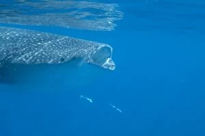
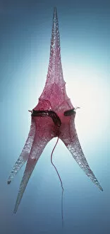
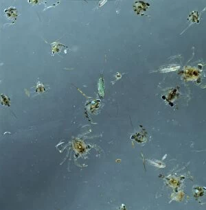
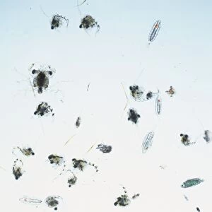
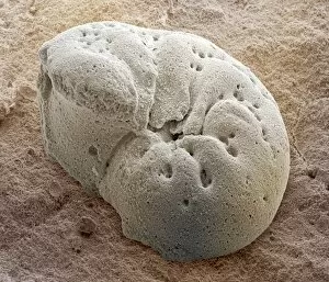
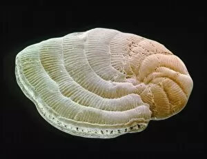
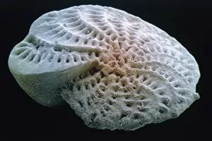
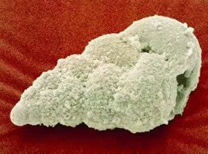
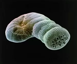
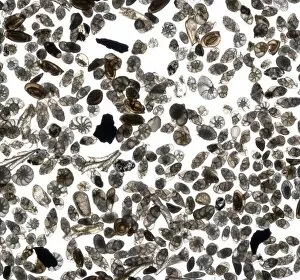
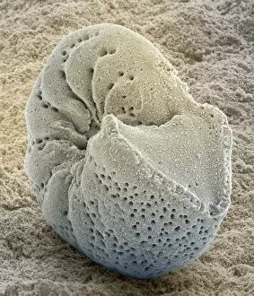
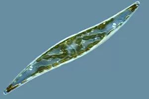
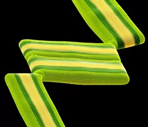
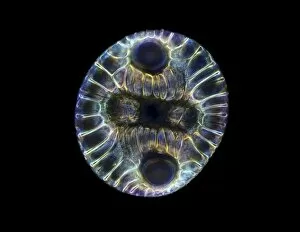
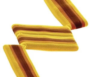
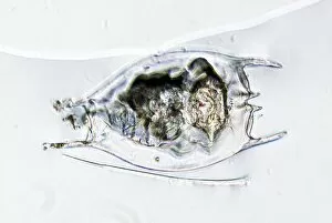
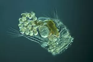
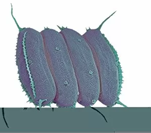
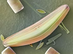
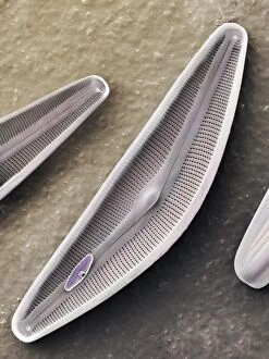
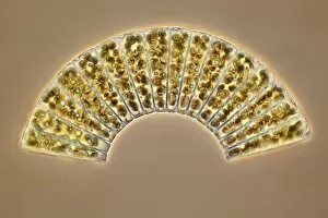
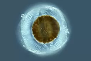
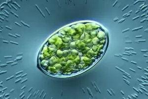
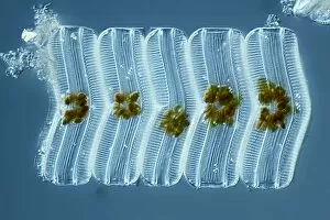
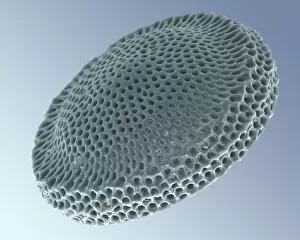
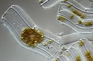
Plankton: Unveiling the Hidden Marvels of the Ocean Dive into the mesmerizing world of plankton, where beauty and wonder intertwine in a delicate dance. From the intricate artistry of diatom algae to the awe-inspiring sight of a whale shark feeding with its mouth wide open, these tiny organisms hold immense significance in our vast oceans. Ernst Haeckel's stunning depiction of diatom algae showcases their exquisite patterns and shapes, reminding us that even microscopic life forms can be true works of art. These diatoms play a crucial role as primary producers, contributing to almost half of Earth's oxygen production. Imagine being a diver off Australia's coast, witnessing firsthand the majestic encounter between a whale shark and its planktonic feast. This gentle giant glides through the water effortlessly, creating an ethereal spectacle that captivates divers from around the world. Examining diatoms under scanning electron microscopy reveals their intricate structures up close. Each detail unravels another layer of complexity within these minuscule organisms, highlighting their importance as essential components in marine ecosystems. The Isle of Man becomes home to basking sharks like Certorhinus maximus during certain times of year. Douglas David Seifert's photograph captures one such moment when this magnificent creature graces us with its presence. Its sheer size reminds us how vital plankton is for sustaining these gentle giants' colossal appetite. In every drop sampled from marine environments lies an entire universe waiting to be explored - each diatom telling its unique story. Through SEM imagery, we gain insight into their diverse forms and functions; they are truly nature's architects shaping our oceans' delicate balance. Returning once again to Cenderawasih Bay in West Papua Indonesia brings yet another breathtaking encounter with whale sharks – Rhincodon typus – gracefully swimming amidst clouds of planktonic abundance. Their presence serves as a reminder that protecting these fragile ecosystems is crucial for the survival of these magnificent creatures.