Plankton Collection (#9)

Plankton: Unveiling the Hidden Marvels of the Ocean Dive into the mesmerizing world of plankton, where beauty and wonder intertwine in a delicate dance

Background imagePlankton Collection: Foraminiferan remains

Foraminiferan remains from the White Cliffs of Dover, U.K. The cliffs are made up of unimaginable numbers of chalky shells of long dead marine animals

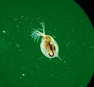
Background imagePlankton Collection: Daphnia, water flea

Daphnia, water flea
A close-up of a water flea (Daphnia sp.), a tiny planktonic crustacean

Background imagePlankton Collection: Pontosphaera japonica

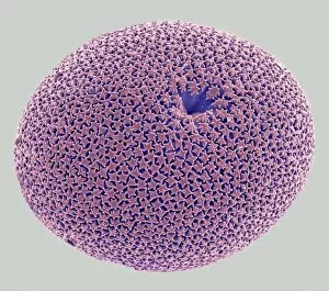
Pontosphaera japonica. A coccolithophore with relatively large, flat, coccoliths. Collected from off Hawaii. Specimen diameter 22m. False-coloured SEM image

Background imagePlankton Collection: Axopodorhabdus albianus, coccolith

Axopodorhabdus albianus, coccolith
Scanning electron microscope image of a Cretaceous coccolith from Folkestone Chalk (x 10, 000 on a standard 9 cm wide print)

Background imagePlankton Collection: Native necklace of fossil foraminifera (Alveolinae)

Native necklace of fossil foraminifera (Alveolinae)
Native necklace of fossil foraminifera from John Whittakers cabinet, fossils dating from the Eocene Limestone, Sindh, India

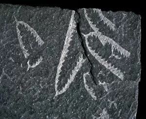
Background imagePlankton Collection: Didymograptus, graptolite

Didymograptus, graptolite
Didymograptus, Ordovician tuning-fork planktonic graptolites. Graptolites are an extinct group of marine, colonial animals

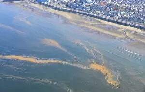
Background imagePlankton Collection: Orange Algae, aerial view of bloom floating on surface of sea, Morecambe Bay, Blackpool

Orange Algae, aerial view of bloom floating on surface of sea, Morecambe Bay, Blackpool, Lancashire, England, August

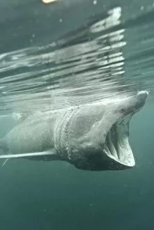
Background imagePlankton Collection: Basking shark (Cetorhinus maximus) feeding on plankton, Hebrides, Scotland, United Kingdom, Europe

Basking shark (Cetorhinus maximus) feeding on plankton, Hebrides, Scotland, United Kingdom, Europe

Background imagePlankton Collection: Marine Diatoms and Radiolarians

Marine Diatoms and Radiolarians
LRMC-17 Scanning Electron Micrograph (SEM): Marine Diatoms and Radiolaria skeletons Magnification x435 (when printed A4, 29.7 cm cm wide)

Background imagePlankton Collection: Moon Jellyfish - Children looking at jellyfish - Monterey Bay Aquarium - CA - *Captive

Moon Jellyfish - Children looking at jellyfish - Monterey Bay Aquarium - CA - *Captive
SE-1776 Moon Jellyfish - Children looking at jellyfish Monterey Bay Aquarium - California Aurelia aurita Monterey Bay Aquarium - California Suzi Eszterhas contact details: prints@ardea.com tel

Background imagePlankton Collection: Moon Jellyfish - Monterey Bay Aquarium - CA - *Captive

Moon Jellyfish - Monterey Bay Aquarium - CA - *Captive
SE-1777 Moon Jellyfish Monterey Bay Aquarium - California Aurelia aurita Monterey Bay Aquarium - California Suzi Eszterhas contact details: prints@ardea.com tel: +44 (0) 20 8318 1401

Background imagePlankton Collection: Sea Nettle - Visitor photographing jellyfish - Monterey Bay Aquarium - CA - *Captive

Sea Nettle - Visitor photographing jellyfish - Monterey Bay Aquarium - CA - *Captive
SE-1778 Sea Nettle - Visitor photographing jellyfish Monterey Bay Aquarium - California Chrysaora fuscescens Monterey Bay Aquarium - California Suzi Eszterhas contact details: prints@ardea.com tel

Background imagePlankton Collection: Mobula Ray - feeding on plankton at the water's surface - Baja CA - Mexico

Mobula Ray - feeding on plankton at the water's surface - Baja CA - Mexico
SE-1304 Mobula Ray - feeding on plankton at the water's surface Baja CA - Mexico Suzi Eszterhas contact details: prints@ardea.com tel: +44 (0) 20 8318 1401

Background imagePlankton Collection: Amoeba - Phase contrast

Amoeba - Phase contrast
JC-183 AMOEBA - PHASE CONTRAST John Clegg contact details: prints@ardea.com tel: +44 (0) 20 8318 1401

Background imagePlankton Collection: Protozoa - x 50 magnification

Protozoa - x 50 magnification
JC-202 Protozoa - x 50 magnification Euglena flagellata John Clegg contact details: prints@ardea.com tel: +44 (0) 20 8318 1401

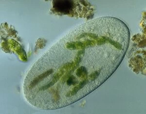
Background imagePlankton Collection: Light Micrograph: Ciliate - Magnification x 750 (when printed A4, 29. 7 cm wide)

Light Micrograph: Ciliate - Magnification x 750 (when printed A4, 29. 7 cm wide)
LRDS-315 Light Micrograph: Ciliate Magnification x 750 (when printed A4, 29.7 cm wide) Frontonia sp. David Spears (Last Refuge) / ardea.com Last Refuge contact details: prints@ardea.com tel

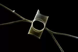
Background imagePlankton Collection: Marine Diatom

Marine Diatom
LRDS-293 Scanning Electron Micrograph (SEM): Marine Diatom Magnification x2, 570 (when printed 10.5 cm wide) Bacteriastrum sp David Spears (Last Refuge) / ardea.com Last Refuge contact details

Background imagePlankton Collection: Marine Diatom

Marine Diatom
LRDS-291 Scanning Electron Micrograph (SEM): Marine Diatom Magnification x1, 510 (when printed 10.5 cm wide) David Spears (Last Refuge) / ardea.com Last Refuge contact details: prints@ardea.com tel

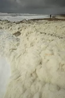
Background imagePlankton Collection: Sea foam washing up on beach after storm, Hengistbury Head, Dorset, England, april

Sea foam washing up on beach after storm, Hengistbury Head, Dorset, England, april

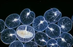
Background imagePlankton Collection: Pl ton -(Noctiluca scintillans) group / one with ingested copepod / x12

Pl ton -(Noctiluca scintillans) group / one with ingested copepod / x12

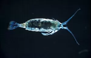
Background imagePlankton Collection: Anomalocera patersoni, female from the marine plankton. A large copepod common at the seas surface

Anomalocera patersoni, female from the marine plankton. A large copepod common at the seas surface in the summer

Background imagePlankton Collection: acrinoptychus senarius living in the plankton x130 magnification

acrinoptychus senarius living in the plankton x130 magnification

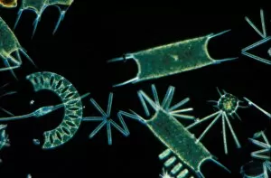
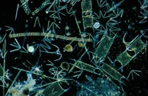
Background imagePlankton Collection: Plankton-Phytopl'ton Phytoplankton x 30 / various diatoms / dino-flagellate etc / English Channel

Plankton-Phytopl'ton Phytoplankton x 30 / various diatoms / dino-flagellate etc / English Channel

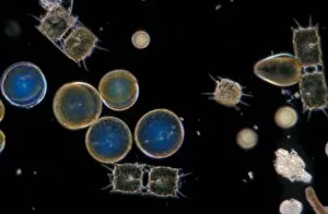
Background imagePlankton Collection: Plankton - Dinoflagellate (Peridinium depressum) x 43

Plankton - Dinoflagellate (Peridinium depressum) x 43

Background imagePlankton Collection: Plankton - Phytoplankton / Winter / x30 / coscinodiscus grani /Biddulphia regia

Plankton - Phytoplankton / Winter / x30 / coscinodiscus grani /Biddulphia regia

Background imagePlankton Collection: Plankton - x 30 Phytoplankton: Various species of diatom eps. Biddilphia sinensis

Plankton - x 30 Phytoplankton: Various species of diatom eps. Biddilphia sinensis

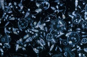
Background imagePlankton Collection: Plankton-Zooplankton Autumn. showing various copepods, cirupede nauplii and obelia medusal. FL006800

Plankton-Zooplankton Autumn. showing various copepods, cirupede nauplii and obelia medusal. FL006800
Plankton-Zooplankton Autumn.showing various copepods, cirupede nauplii and obelia medusal. FL006800

Background imagePlankton Collection: Plankton - Diatom (Rhizosolenia stolterfothii) Individual diatoms unite to form

Plankton - Diatom (Rhizosolenia stolterfothii) Individual diatoms unite to form

Background imagePlankton Collection: Plankton-Zooplankton (Autumn) Cofepods (Calanus, Centro- pages, Acartia, Temora etc. )

Plankton-Zooplankton (Autumn) Cofepods (Calanus, Centro- pages, Acartia, Temora etc. )
Plankton-Zooplankton (Autumn) Cofepods (Calanus, Centro- pages, Acartia, Temora etc.)

Background imagePlankton Collection: Plankton-Phytopl'ton X20 / various species of diatom & dinoflagellates / English Channel

Plankton-Phytopl'ton X20 / various species of diatom & dinoflagellates / English Channel

Background imagePlankton Collection: Ciliate protozoa, light micrograph

Ciliate protozoa, light micrograph
Ciliate protozoa. Light micrograph of a mixed population of freshwater ciliate protozoa and algae. The algae are the green filaments crossing the field

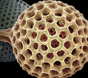
Background imagePlankton Collection: Radiolarian planktonic protozoan, SEM

Radiolarian planktonic protozoan, SEM
Radiolarian protozoan. Coloured scanning electron micrograph (SEM) of the shell (test) of an unidentified radiolarian protozoan

Background imagePlankton Collection: Orbulina foraminiferan, SEM

Orbulina foraminiferan, SEM
Orbulina. Coloured scanning electron micrograph (SEM) of the shell of the foraminiferan Orbulina sp. Foraminiferans are marine single-celled protists that construct and inhabit shells (tests)

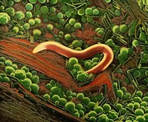
Background imagePlankton Collection: False col SEM of nematode worm on sample of peat

False col SEM of nematode worm on sample of peat
False-colour scanning electron micrograph of a nematode worm on a sample of peat. The micrograph shows the layer of " green slime" which often appears on peat & other organic surfaces

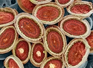
Background imagePlankton Collection: Plankton cell wall, SEM

Plankton cell wall, SEM
Plankton cell wall. Coloured scanning electron micrograph (SEM) of the geometrical plates making up the mineralised cell wall of a planktonic alga (Coronosphaera mediterranea)

Background imagePlankton Collection: Krill head, SEM

Krill head, SEM
Antarctic krill head (Euphausia superba), coloured scanning electron micrograph (SEM). This animal is a marine crustacean and an important food for many marine animals

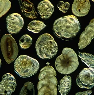
Background imagePlankton Collection: LM of assorted fossil Foraminifera shells

LM of assorted fossil Foraminifera shells
Foraminiferan shells. Light micrograph of assorted shells of fossil species belonging to the order: Foraminifera. Single-celled protozoans once inhabited these shells which are composed of several

Background imagePlankton Collection: Rotifer worm, light micrograph

Rotifer worm, light micrograph
Rotifer worm. Light micrograph of the rotifer worm Platyas quadricornis. This free-swimming, aquatic micro-organism has a body which is divided into three sections

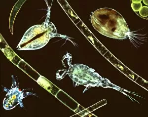
Background imagePlankton Collection: Aquatic crustaceans, light micrograph

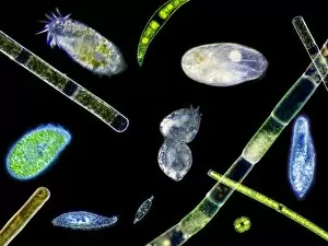
Aquatic crustaceans, light micrograph
Aquatic crustaceans. Composite image of light micrographs of aquatic planktonic crustaceans with strands of filamentous algae

Background imagePlankton Collection: Lychnocanium radiolarian, SEM

Lychnocanium radiolarian, SEM
Lychnocanium radiolarian. Coloured scanning electron micrograph (SEM) of the shell of a Lychnocanium sp. radiolarian. Radiolaria are single-celled protozoans that are found in marine plankton

Background imagePlankton Collection: LM of copepods, marine crustaceans in plankton

LM of copepods, marine crustaceans in plankton
Copepods. Light micrograph of a group of copepods, subclass Copepoda, marine crustaceans. Copepods are tiny, shrimp-like planktonic animals that live in vast numbers in the surface of seas and lakes

Background imagePlankton Collection: Amphisphaerina radiolarian, SEM

Amphisphaerina radiolarian, SEM
Amphisphaerina radiolarian. Coloured scanning electron micrograph (SEM) of the shell of a Amphisphaerina sp. radiolarian. Radiolaria are single-celled protozoans that are found in marine plankton

Background imagePlankton Collection: Acrosphaera radiolarian, SEM

Acrosphaera radiolarian, SEM
Acrosphaera radiolarian. Coloured scanning electron micrograph (SEM) of the shell of a Acrosphaera sp. radiolarian. Radiolaria are single-celled protozoans that are found in marine plankton

Background imagePlankton Collection: LM of assorted Foraminifera shells

LM of assorted Foraminifera shells
Foraminiferan shells. Light micrograph of assorted species of shells belonging to the order Foramini- fera. Single-celled protozoans inhabit these shells which are composed of several chambers

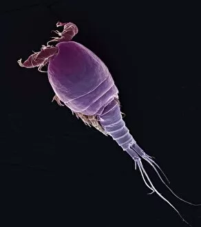
Background imagePlankton Collection: Cyclops sp. copepod, SEM

Cyclops sp. copepod, SEM
Cyclops sp. copepod. Coloured scanning electron micrograph (SEM) of a freshwater copepod (Cyclops sp.). The head of the animal is at upper left


For sale as Licensed Images

Choose your image, Select your licence and Download the media

Plankton: Unveiling the Hidden Marvels of the Ocean Dive into the mesmerizing world of plankton, where beauty and wonder intertwine in a delicate dance. From the intricate artistry of diatom algae to the awe-inspiring sight of a whale shark feeding with its mouth wide open, these tiny organisms hold immense significance in our vast oceans. Ernst Haeckel's stunning depiction of diatom algae showcases their exquisite patterns and shapes, reminding us that even microscopic life forms can be true works of art. These diatoms play a crucial role as primary producers, contributing to almost half of Earth's oxygen production. Imagine being a diver off Australia's coast, witnessing firsthand the majestic encounter between a whale shark and its planktonic feast. This gentle giant glides through the water effortlessly, creating an ethereal spectacle that captivates divers from around the world. Examining diatoms under scanning electron microscopy reveals their intricate structures up close. Each detail unravels another layer of complexity within these minuscule organisms, highlighting their importance as essential components in marine ecosystems. The Isle of Man becomes home to basking sharks like Certorhinus maximus during certain times of year. Douglas David Seifert's photograph captures one such moment when this magnificent creature graces us with its presence. Its sheer size reminds us how vital plankton is for sustaining these gentle giants' colossal appetite. In every drop sampled from marine environments lies an entire universe waiting to be explored - each diatom telling its unique story. Through SEM imagery, we gain insight into their diverse forms and functions; they are truly nature's architects shaping our oceans' delicate balance. Returning once again to Cenderawasih Bay in West Papua Indonesia brings yet another breathtaking encounter with whale sharks – Rhincodon typus – gracefully swimming amidst clouds of planktonic abundance. Their presence serves as a reminder that protecting these fragile ecosystems is crucial for the survival of these magnificent creatures.