
Programmed Cell Death Collection
"Unveiling the Intricacies of Programmed Cell Death: A Fascinating Journey into Cellular Self-Destruction" In this captivating journey


For sale as Licensed Images
Choose your image, Select your licence and Download the media
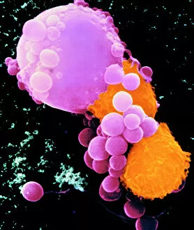
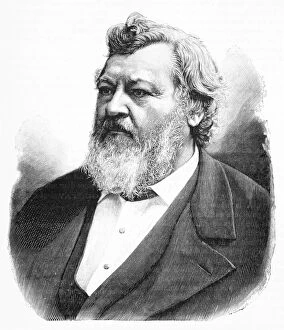
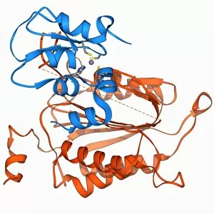
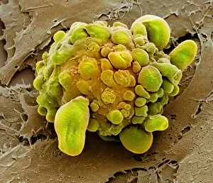
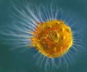
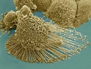
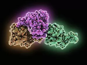
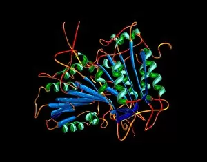
"Unveiling the Intricacies of Programmed Cell Death: A Fascinating Journey into Cellular Self-Destruction" In this captivating journey, we explore the phenomenon of programmed cell death, a fundamental process that safeguards our bodies against diseases and maintains cellular balance. Through a mesmerizing colored scanning electron microscope (SEM) image, we witness lymphocytes fearlessly launching an attack on cancer cells. This powerful visual reminds us of the intricate mechanisms at play within our immune system. We delve into history as we pay homage to Carl Vogt, the visionary German naturalist who first proposed the concept in 1842. His groundbreaking insights laid the foundation for future discoveries in this field. Zooming closer into molecular intricacies, we encounter two key players: Programmed Cell Death Protein Molecule F006/9597 and Programmed Cell Death Protein 6 Molecule F006/9548. These proteins orchestrate cellular demise with precision and finesse, ensuring proper elimination without causing harm to surrounding healthy tissues. Transcription factor and DNA molecule F006/9484 come together in perfect harmony to regulate gene expression during programmed cell death. Their synchronized dance ensures that only specific genes are activated or silenced when it is time for a cell to meet its fate. Meet Caspase-3 and its inhibitor F006/9457 - an elegant duo responsible for orchestrating apoptosis by cleaving vital cellular components. Together they maintain a delicate balance between life and death within our cells. Caspase-9 takes center stage alongside its inhibitor in a striking molecular model F006/9442. This dynamic pair acts as gatekeepers, controlling whether cells succumb to their destiny or evade destruction altogether. Witnessing apoptosis firsthand through transmission electron microscopy (TEM), we observe cells undergoing controlled self-destruction while maintaining their structural integrity—a testament to nature's remarkable design. Even stem cells are not exempt from programmed cell death's reach, as shown through multiple scanning electron microscope (SEM) images.