Protozoa Collection (#6)

Protozoa, the microscopic wonders of nature

Background imageProtozoa Collection: Foraminifera

Foraminifera
Part of the display of foraminifera from The Great Exhibition of 1851. Featured are specimens from the London Clay, the Paris Basin and the Gulf of Suez

Background imageProtozoa Collection: Difflugia Corona

Difflugia Corona
Freshwater Testate Amoebae. Magnification x 450

Background imageProtozoa Collection: Trypanosomes

Trypanosomes
Scanning electron microscope image showing a trypanosoma blood smear. They have proved to be of great interest as they have evolved very differently to other better studied organisms

Background imageProtozoa Collection: Myxomycetes, plasmodial slime mould

Myxomycetes, plasmodial slime mould
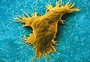
Scanning electron microscope image of a plasmodial slime mould spore (x12000). This mould spends most of its life as a single cell; when they reproduce they form a slug-like blob that can travel

Background imageProtozoa Collection: Acanthowetra

Acanthowetra
A photograph of a foraminifera found in the Indian Ocean

Background imageProtozoa Collection: Foraminiferan remains

Foraminiferan remains from the White Cliffs of Dover, U.K. The cliffs are made up of unimaginable numbers of chalky shells of long dead marine animals

Background imageProtozoa Collection: Minakatella longifila, slime mould

Minakatella longifila, slime mould

Background imageProtozoa Collection: Amoeba - Phase contrast

Amoeba - Phase contrast
JC-183 AMOEBA - PHASE CONTRAST John Clegg contact details: prints@ardea.com tel: +44 (0) 20 8318 1401

Background imageProtozoa Collection: Protozoa - x 50 magnification

Protozoa - x 50 magnification
JC-202 Protozoa - x 50 magnification Euglena flagellata John Clegg contact details: prints@ardea.com tel: +44 (0) 20 8318 1401

Background imageProtozoa Collection: Light Micrograph: Ciliate - Magnification x 750 (when printed A4, 29. 7 cm wide)

Light Micrograph: Ciliate - Magnification x 750 (when printed A4, 29. 7 cm wide)
LRDS-315 Light Micrograph: Ciliate Magnification x 750 (when printed A4, 29.7 cm wide) Frontonia sp. David Spears (Last Refuge) / ardea.com Last Refuge contact details: prints@ardea.com tel

Background imageProtozoa Collection: Light Micrograph: Plasmodium: a parasitic protozoa in blood; Magnification x 7

Light Micrograph: Plasmodium: a parasitic protozoa in blood; Magnification x 7, 500 (if print A4 size: 29. 7 cm wide)
LRDS-313 Light Micrograph: Plasmodium: a parasitic protozoa in blood; Magnification x 7, 500 (if print A4 size: 29.7 cm wide) David Spears (Last Refuge) / ardea.com Last Refuge contact details

Background imageProtozoa Collection: Metopus protozoan

Metopus protozoan

Background imageProtozoa Collection: Thalassomyxa australis protozoan

Thalassomyxa australis protozoan
Thalassomyxa protozoan. Coloured scanning electron micrograph (SEM) of a Thalassomyxa australis prot- ozoan (single-celled animal)

Background imageProtozoa Collection: Lembadion protozoan

Lembadion protozoan. Coloured scanning electron micrograph (SEM) of a Lembadion bullinum protozoan (single-celled animal)

Background imageProtozoa Collection: TEM of Giardia lamblia

TEM of Giardia lamblia
Coloured Transmission Electron Micrograph of Giardia lamblia (orange), a parasitic flagellate protozoan which causes the disease lambliasis (giardiasis)

Background imageProtozoa Collection: Ciliate protozoa, light micrograph

Ciliate protozoa, light micrograph
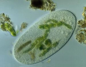
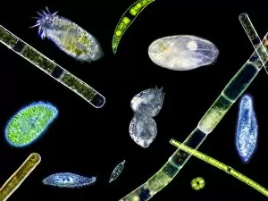
Ciliate protozoa. Light micrograph of a mixed population of freshwater ciliate protozoa and algae. The algae are the green filaments crossing the field

Background imageProtozoa Collection: Radiolarian planktonic protozoan, SEM

Radiolarian planktonic protozoan, SEM
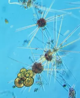
Radiolarian protozoan. Coloured scanning electron micrograph (SEM) of the shell (test) of an unidentified radiolarian protozoan

Background imageProtozoa Collection: Calcareous phytoplankton fossil, artwork

Calcareous phytoplankton fossil, artwork
Calcareous phytoplankton fossil. Artwork of a fossil of the skeleton (coccosphere) of a coccolithotrope, a small marine algal organism

Background imageProtozoa Collection: Leishmania protozoa

Leishmania protozoa
False-colour light micrograph (LM) of a colony of Leishmania tropica. a parasitic flagellate protozoan that causes the disease leishmaniasis in humans

Background imageProtozoa Collection: Ciliate protozoa dividing

Ciliate protozoa dividing
Ciliate protozoa. Light micrograph of unidentified ciliate protozoa (class Ciliata) undergoing asexual reproduction. These unicellular micro-organisms can multiply by either of two methods

Background imageProtozoa Collection: Euplotes ciliate protozoan, SEM

Euplotes ciliate protozoan, SEM
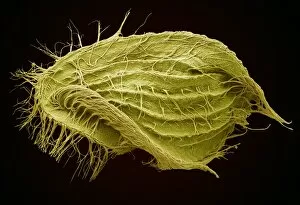
Euplotes ciliate protozoan. Coloured scanning electron micrograph (SEM) of a ciliate protozoan (Euplotes sp.). This single-celled organism is found in fresh and marine water

Background imageProtozoa Collection: Plankton cell wall, SEM

Plankton cell wall, SEM
Plankton cell wall. Coloured scanning electron micrograph (SEM) of the geometrical plates making up the mineralised cell wall of a planktonic alga (Coronosphaera mediterranea)

Background imageProtozoa Collection: Spirochona protozoa

Spirochona protozoa

Background imageProtozoa Collection: Euglena protozoan, SEM

Euglena protozoan, SEM
Euglena protozoan (Euglena sp.), coloured scanning electron micrograph (SEM). This protozoan is described as flagellate because it has a flagellum (thin tail-like structure)

Background imageProtozoa Collection: LM of assorted fossil Foraminifera shells

LM of assorted fossil Foraminifera shells
Foraminiferan shells. Light micrograph of assorted shells of fossil species belonging to the order: Foraminifera. Single-celled protozoans once inhabited these shells which are composed of several

Background imageProtozoa Collection: Protozoan, SEM

Protozoan, SEM
Protozoan. Coloured scanning electron micrograph (SEM) of a protozoan. Protozoa are unicellular eukaryotic organisms, that usually are not photosynthetic

Background imageProtozoa Collection: Ciliate protozoans, SEM

Ciliate protozoans, SEM
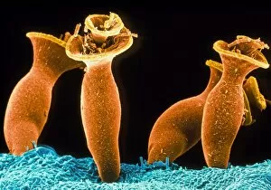
Ciliate protozoans. Coloured scanning electron micrograph (SEM) of sessile (anchored) ciliate protozoan (subclass Peritrichia)

Background imageProtozoa Collection: Giardia protozoan, TEM

Giardia protozoan, TEM
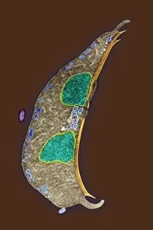
Giardia protozoan. Coloured transmission electron micrograph (TEM) of a Giardia sp. protozoan. It is a single-celled organism that has two cell nuclei (green)

Background imageProtozoa Collection: Calcareous phytoplankton plates, SEM

Calcareous phytoplankton plates, SEM
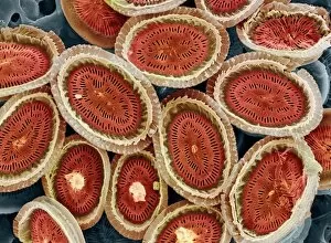
Calcareous phytoplankton plates. Coloured scanning electron micrograph (SEM) of parts of the skeleton (coccosphere) of the coccolithophore Emiliania huxleyi, a small marine algal organism

Background imageProtozoa Collection: Trichomytopsis protozoan

Trichomytopsis protozoan

Background imageProtozoa Collection: Coloured SEM of Giardia lamblia in human intestine

Coloured SEM of Giardia lamblia in human intestine
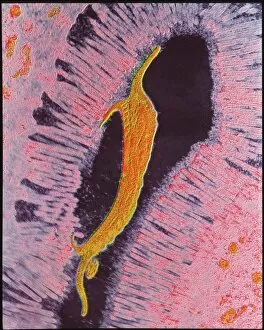
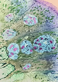
Giardia lamblia. Coloured Scanning Electron Micro- graph (SEM) of the parasite Giardia lamblia in the human small intestine

Background imageProtozoa Collection: Ciliate protozoa, SEM

Ciliate protozoa, SEM
Ciliate protozoa. Coloured scanning electron micrograph (SEM) of cilliate protozoa (unicellular animals). The ciliates are so named because of the hair-like cilia surrounding their bodies which are

Background imageProtozoa Collection: LM of a colony of Volvox

LM of a colony of Volvox
Light micrograph of a colony of Volvox containing eight asexual daughter colonies within. Volvox is an organism that lies between the plant & animal kingdom

Background imageProtozoa Collection: Naegleria fowleri protozoa, TEM

Naegleria fowleri protozoa, TEM
Naegleria fowleri protozoan. Coloured transmission electron micrograph (TEM) of a section through a Naegleria fowleri protozoan

Background imageProtozoa Collection: Flagellate protozoan, SEM

Flagellate protozoan, SEM
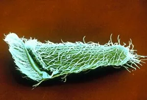
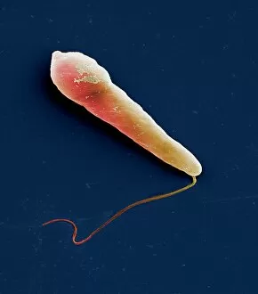
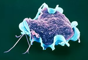
Flagellate protozoan, coloured scanning electron micrograph (SEM). This is a protozoan that has a flagellum (thin tail-like structure, lower left)

Background imageProtozoa Collection: Parasitic protozoan, TEM

Parasitic protozoan, TEM
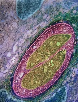
Parasitic protozoan. Coloured transmission electron micrograph (TEM) of a trypanosome protozoan (Trypanosoma sp.). The nucleus (centre), cell membrane (outline)

Background imageProtozoa Collection: Enterocytozoon sp. parasites, TEM

Enterocytozoon sp. parasites, TEM

Background imageProtozoa Collection: Isospora sp. parasites, TEM

Isospora sp. parasites, TEM
Isospora sp. parasites. Coloured transmission electron micrograph (TEM) of Isospora sp. parasites (yellow) in a sectioned cell

Background imageProtozoa Collection: Lychnocanium radiolarian, SEM

Lychnocanium radiolarian, SEM
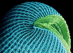
Lychnocanium radiolarian. Coloured scanning electron micrograph (SEM) of the shell of a Lychnocanium sp. radiolarian. Radiolaria are single-celled protozoans that are found in marine plankton

Background imageProtozoa Collection: TEM of Leishmania sp

TEM of Leishmania sp
False-colour transmission electron micrograph (TEM) of Leishmania sp. a parasitic flagellate protozoan that causes the disease leishmaniasis in humans


For sale as Licensed Images

Choose your image, Select your licence and Download the media

Protozoa, the microscopic wonders of nature. From calcareous phytoplankton fossils to intricate artwork depicting Trypanosome protozoans, these single-celled organisms never cease to amaze. Take a closer look through the lens of SEM Z100 / 0213 and discover the mesmerizing beauty of their world. In one frame, we witness Plasmodium sp. , a malarial parasite that wreaks havoc on human health. Its complex structure reminds us of the delicate balance between life and disease. But not all it can harmful; many lead fascinating lives as scavengers or nutrient absorbers. Through light micrograph LM, we observe a kidney-shaped ciliate surrounded by Euglena sp. , showcasing their feeding habits at an astonishing magnification of x900. Diatoms, another group of protozoa, steal our attention with their intricate patterns and shapes. Acrosphaera radiolarian captivates us in its SEM image, displaying its ornate exoskeleton that protects it from predators. Foraminiferan tests (shells) take center stage in another SEM capture. These tiny structures tell stories about ancient oceans and provide valuable insights into Earth's history. Moving away from imagery, we encounter models representing foraminifera - miniature replicas that aid scientists in understanding these remarkable creatures' behavior and evolution. Not limited to aquatic environments alone, some protozoa find residence within our own bodies as intestinal parasites. TEM images reveal their presence while reminding us to prioritize hygiene and health practices. Amongst them is Trichomonas vaginalis captured through TEM - a reminder that even seemingly harmless organisms can cause discomfort if left unchecked. Lastly, Cryptosporidium protozoa appear under TEM's watchful eye – highlighting the importance of water safety measures due to this organism's ability to contaminate drinking sources with potential health risks. Protozoa may be small in size, but their impact on our world is immense.