
Protozoa Collection (#2)
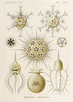
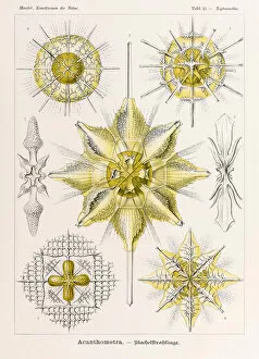
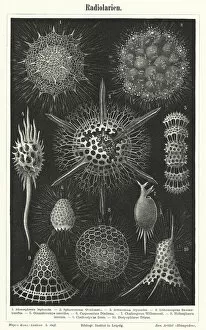
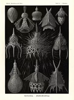
Protozoa, the microscopic wonders of nature


For sale as Licensed Images
Choose your image, Select your licence and Download the media
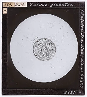
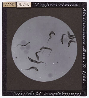
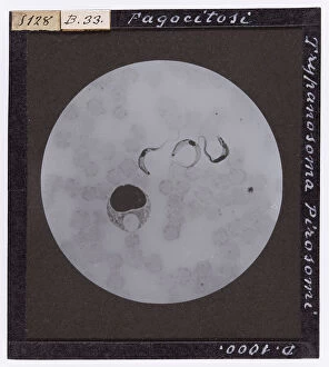
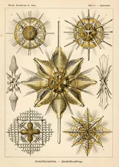
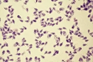
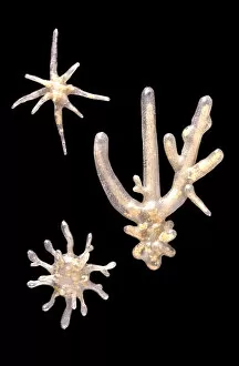
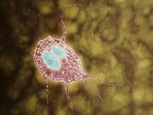
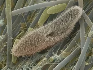
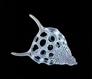
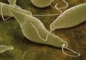
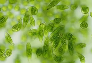
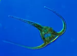
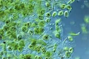
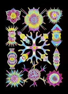
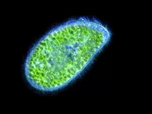
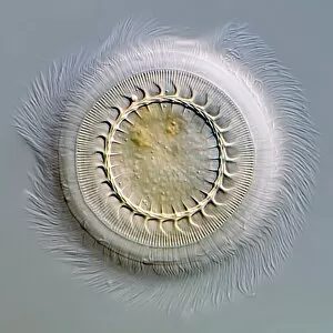
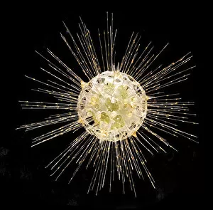
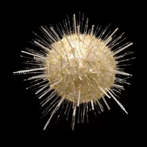
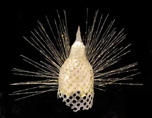
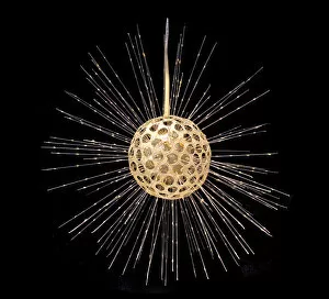
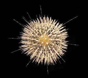
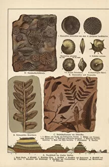
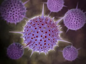
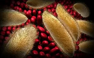
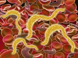
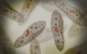
Protozoa, the microscopic wonders of nature. From calcareous phytoplankton fossils to intricate artwork depicting Trypanosome protozoans, these single-celled organisms never cease to amaze. Take a closer look through the lens of SEM Z100 / 0213 and discover the mesmerizing beauty of their world. In one frame, we witness Plasmodium sp. , a malarial parasite that wreaks havoc on human health. Its complex structure reminds us of the delicate balance between life and disease. But not all it can harmful; many lead fascinating lives as scavengers or nutrient absorbers. Through light micrograph LM, we observe a kidney-shaped ciliate surrounded by Euglena sp. , showcasing their feeding habits at an astonishing magnification of x900. Diatoms, another group of protozoa, steal our attention with their intricate patterns and shapes. Acrosphaera radiolarian captivates us in its SEM image, displaying its ornate exoskeleton that protects it from predators. Foraminiferan tests (shells) take center stage in another SEM capture. These tiny structures tell stories about ancient oceans and provide valuable insights into Earth's history. Moving away from imagery, we encounter models representing foraminifera - miniature replicas that aid scientists in understanding these remarkable creatures' behavior and evolution. Not limited to aquatic environments alone, some protozoa find residence within our own bodies as intestinal parasites. TEM images reveal their presence while reminding us to prioritize hygiene and health practices. Amongst them is Trichomonas vaginalis captured through TEM - a reminder that even seemingly harmless organisms can cause discomfort if left unchecked. Lastly, Cryptosporidium protozoa appear under TEM's watchful eye – highlighting the importance of water safety measures due to this organism's ability to contaminate drinking sources with potential health risks. Protozoa may be small in size, but their impact on our world is immense.