Pulmonary Collection (page 8)

"Pulmonary: Unveiling the Intricacies of our Vital Breath" Discover the wonders of the pulmonary system through a captivating journey

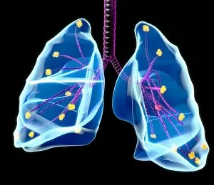
Background imagePulmonary Collection: Human lungs, artwork

Human lungs, artwork
Human lungs, computer artwork

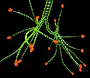
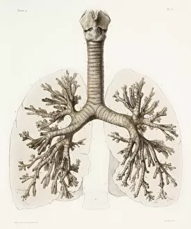
Background imagePulmonary Collection: Bronchial tree, artwork

Bronchial tree, artwork
Bronchial tree, computer artwork

Background imagePulmonary Collection: TB bacteria infecting macrophages, SEM

TB bacteria infecting macrophages, SEM
TB bacteria infecting macrophages. Coloured scanning electron micrograph (SEM) of Mycobacterium tuberculosis bacteria (orange) invading macrophage white blood cells (pink)

Background imagePulmonary Collection: Alveoli in the lung, artwork

Alveoli in the lung, artwork
Alveoli in the lung. Computer artwork showing the alveoli (spheres) and bronchioles (tubes) of the lungs. The alveoli are the tiny air sacs of the lungs where gases are exchanged between the air in

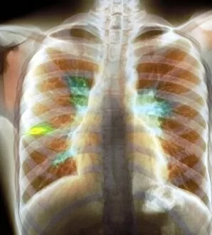
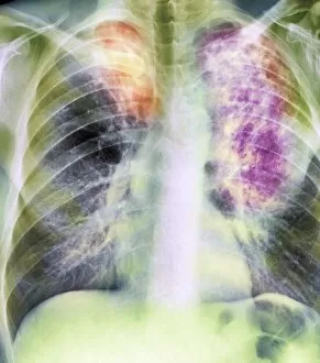
Background imagePulmonary Collection: Collapse (atelectasis) of lung, X-ray

Collapse (atelectasis) of lung, X-ray
Coloured X-ray (front view) of the chest in a 58 year old patient showing collapse (atelectasis) of part of the upper lobe of a lung (orange, at left)

Background imagePulmonary Collection: Lung abscess, CT scan

Lung abscess, CT scan
Coloured computed tomography (CT) scan in axial view of the lung fields in a 47 year old patient showing the presence of an abscess (black) in each lung

Background imagePulmonary Collection: Pulmonary lobule, artwork

Pulmonary lobule, artwork
Pulmonary lobule. Computer artwork of a section through an area of lung tissue. A pulmonary lobule is a unit of lung tissue that includes a bronchiole, alveolar ducts, sacs, and alveoli

Background imagePulmonary Collection: Tuberculosis diagnosis, 19th century

Tuberculosis diagnosis, 19th century. Artwork showing the French physician Rene Laennec (1781-1826), at the Necker Hospital, Paris, France, examining a patient (left)

Background imagePulmonary Collection: Tuberculosis lymphadenopathy, X-ray

Tuberculosis lymphadenopathy, X-ray
Coloured X-ray (front view) of the chest in a 14 year old male patient, showing inflamed lymph nodes (lymphadenopathy, yellow, at centre right and left) caused by tuberculosis (TB)

Background imagePulmonary Collection: Asthma inhaler use, artwork

Asthma inhaler use, artwork
Asthma inhale use. Artwork showing how to use an asthma inhaler. Asthma is a condition in which the small airways (bronchioles) in the lungs constrict, making it difficult to breathe

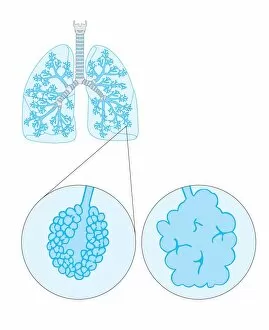
Background imagePulmonary Collection: Emphysema, artwork

Emphysema, artwork
Emphysema. Artwork showing the lungs with a detailed view of a normal alveolus (air sac, left) and an alveolus affected by emphysema (right)

Background imagePulmonary Collection: Pulmonary consolidation, X-ray

Pulmonary consolidation, X-ray
Pulmonary consolidation. X-ray showing that the upper lobe of a patients left lung has become consolidated (filled with pus) due to pneumonia

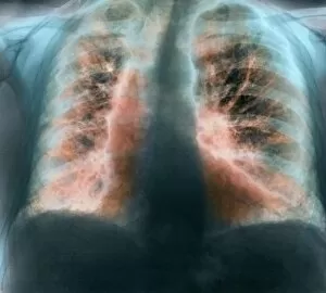
Background imagePulmonary Collection: Old and new tuberculosis, X-ray

Old and new tuberculosis, X-ray
Coloured X-ray (front view) of the chest in a 50 year old male patient, showing fibrosis in the lungs from old tuberculosis (more fibrous scarring, orange)

Background imagePulmonary Collection: Heart and lungs, historical illustration

Heart and lungs, historical illustration. 19th Century hand coloured lithographic print showing the lungs (left and right) situated either side of the heart (lower centre)

Background imagePulmonary Collection: Respiratory anatomy, 19th Century artwork

Respiratory anatomy, 19th Century artwork. Historical hand coloured lithographic print showing the trachea (wind pipe, vertical) running down from the larynx (voicebox)

Background imagePulmonary Collection: Heart and neck, historical illustration

Heart and neck, historical illustration. 19th Century hand coloured lithographic print showing the major blood vessels (red and blue) and nerves (white) that pass through the human neck

Background imagePulmonary Collection: Fibrosing alveolitis, X-ray

Fibrosing alveolitis, X-ray
Fibrosing alveolitis. Coloured X-ray of the chest of an 82-year-old man with fibrosing alveolitis. Also known as idiopathic pulmonary fibrosis and cryptogenic fibrosing alveolitis

Background imagePulmonary Collection: Respiratory nerves, 1844 artwork

Respiratory nerves, 1844 artwork
Respiratory nerves. These anatomical artworks are figures 2-3, plate 49, volume 3 (1844) of Traite complet de l anatomie de l homme (1831-1854)

Background imagePulmonary Collection: Lung cells, fluorescent micrograph

Lung cells, fluorescent micrograph
Lung cells. Immunofluorescence light micrograph of pulmonary endothelial cells. Endothelial cells are specialized epithelial cells that line the inner surface of blood vessels

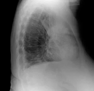
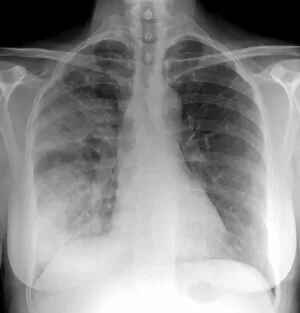
Background imagePulmonary Collection: Pneumonia, X-ray

Pneumonia, X-ray

Background imagePulmonary Collection: Lung alveolus, artwork

Lung alveolus, artwork
Lung alveolus, computer artwork. Alveoli are the site of gas exchange in the lungs of mammals, where oxygen is taken up and carbon dioxide released

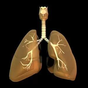
Background imagePulmonary Collection: Human lungs

Human lungs. Anatomical artwork of the human lungs and respiratory system. The heart (which has been removed here) is located between the lungs

Background imagePulmonary Collection: Airways of the lungs, artwork

Airways of the lungs, artwork
Artwork of the airways of the lungs showing the trachea (windpipe, at top) dividing into two bronchi which in turn divide into many bronchioles supplying air during breathing (respiration)

Background imagePulmonary Collection: Human coronavirus, TEM

Human coronavirus, TEM
Human coronavirus. Coloured transmission electron micrograph (TEM) of Human coronavirus particles. Coronaviruses primarily infect the upper respiratory and gastrointestinal tract

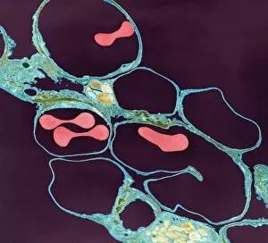
Background imagePulmonary Collection: Lung alveoli and red blood cells, TEM

Lung alveoli and red blood cells, TEM
Lung alveoli and red blood cells. Coloured transmission electron micrograph (TEM) of a section through lung alveoli (blue) and red blood cells (red)

Background imagePulmonary Collection: Lung alveoli and blood cells, TEM

Lung alveoli and blood cells, TEM
Lung alveoli and blood cells. Coloured transmission electron micrograph (TEM) of a section through lung alveoli and blood cells

Background imagePulmonary Collection: Asthma and mucus overproduction, artwork

Asthma and mucus overproduction, artwork
Asthma and mucus overproduction. Artwork showing how the bodys immune response causes the inflammation seen in asthma. The chest of a human figure is at top right

Background imagePulmonary Collection: Lungs and trachea, artwork

Lungs and trachea, artwork. The lungs are where blood is oxygenated. Breathing brings air into the lungs through the trachea (windpipe) at upper centre

Background imagePulmonary Collection: Snake ciliated lung cells and mucus, SEM

Snake ciliated lung cells and mucus, SEM
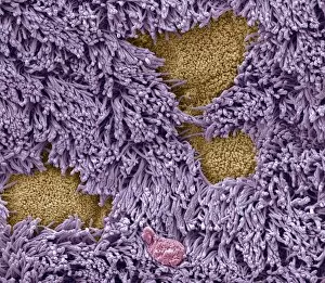
Snake ciliated lung cells and mucus. Coloured scanning electron micrograph (SEM) of ciliated cells (green) and mucus (pink) in the lung of a snake

Background imagePulmonary Collection: Lung bronchus cross-section, SEM

Lung bronchus cross-section, SEM
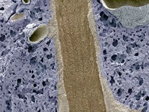
Lung bronchus cross-section, coloured scanning electron micrograph (SEM). This longitudinal freeze-fracture has exposed the inner epithelial surface of a bronchus (brown)

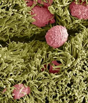
Background imagePulmonary Collection: Nasal epithelium, SEM

Nasal epithelium, SEM
Nasal epithelium, coloured scanning electron micrograph (SEM). The nasal cavity contains both cilated (pink) and non-ciliated (secretory) cells

Background imagePulmonary Collection: Heart and lungs, 3D CT scan

Heart and lungs, 3D CT scan
Heart and lungs, 3D-CT scan. Coloured 3-D computed tomography (CT) scan of the heart (middle centre) and lungs, showing the small airways (bronchioles) of the lungs. The rib cage surrounds the lungs

Background imagePulmonary Collection: Bronchodilator drug crystals, SEM

Bronchodilator drug crystals, SEM
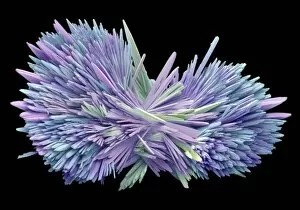
Bronchodilator drug crystals, coloured scanning electron micrograph (SEM). These are crystals of the drug ventolin (salbutamol sulphate), a bronchodilator drug used to treat asthma

Background imagePulmonary Collection: Pneumonia infection, light micrograph

Pneumonia infection, light micrograph
Pneumonia infection. Light micrograph of a section through lung tissue from a patient with bronchial pneumonia. Shown here is the blockage and breakdown of the air sacs

Background imagePulmonary Collection: Artificial heart, conceptual artwork

Artificial heart, conceptual artwork
Artificial heart. Conceptual high-contrast computer artwork of an artificial heart showing its blood supply

Background imagePulmonary Collection: Bronchial tree and lungs, artwork

Bronchial tree and lungs, artwork
Bronchial tree. Computer artwork representing a human bronchial tree and lungs. The human bronchial tree is the network of airways serving both lungs

Background imagePulmonary Collection: Millfield Seaside Home, Littlehampton, Sussex

Millfield Seaside Home, Littlehampton, Sussex
Children and nurses outside the Millfield Seaside Home at Rustington, Littlehampton, Sussex. The Home was opened by the Metropolitan Asylums Board in 1904 as a seaside sanatorium for children

Background imagePulmonary Collection: Sea bathing at Millfield Seaside Home, Littlehampton, Sussex

Sea bathing at Millfield Seaside Home, Littlehampton, Sussex
Sea bathing at the Millfield Seaside Home at Rustington, Littlehampton, Sussex. The Home was opened by the Metropolitan Asylums Board in 1904 as a seaside sanatorium for children suffering


For sale as Licensed Images

Choose your image, Select your licence and Download the media

"Pulmonary: Unveiling the Intricacies of our Vital Breath" Discover the wonders of the pulmonary system through a captivating journey. Dive into a world where life-sustaining breaths take center stage, as we explore various aspects that define our respiratory health. Embark on this educational voyage with a detailed diagram showcasing the intricate structure of lungs and bronchial tubes. Marvel at their complexity, understanding how they facilitate oxygen exchange within our bodies. Witness medical advancements in pneumothorax treatment through X-ray imagery, offering hope to those affected by this condition. Delve into neck anatomy studies, unraveling the secrets hidden beneath our skin while appreciating 19th Century artwork depicting these discoveries. Uncover the devastating impact of tuberculosis through haunting X-ray images that reveal its presence within afflicted lungs. Gain insight into how interconnected systems work together harmoniously with an enlightening diagram illustrating the heart, lungs, and windpipe. Experience tension pneumothorax's urgency via gripping X-rays that depict its severity and prompt intervention requirements. Grasp the symbiotic relationship between heart and lungs; two vital organs intricately linked for optimal functioning. Explore cystic fibrosis' challenges faced by patients daily; understand their resilience amidst adversity. Learn about Konstantin Buteyko's groundbreaking contributions as a Soviet doctor revolutionizing respiratory therapy techniques. Marvel at abdominal arteries' inner workings revealed in an illuminating X-ray image (P206 / 0309), highlighting their significance in maintaining overall health. Discover Hantavirus' impact on pulmonary health – raising awareness about potential risks lurking in nature's realm. Reflect upon "HOCHE DIES AT WETZLAR, " reminding us of historical milestones shaping modern medicine's understanding diseases throughout time. Join us on this extraordinary expedition exploring every facet of "pulmonary. " Let curiosity guide you as we unravel mysteries surrounding respiration - an awe-inspiring process essential to our very existence.