Pulmonary Collection (page 5)

"Pulmonary: Unveiling the Intricacies of our Vital Breath" Discover the wonders of the pulmonary system through a captivating journey

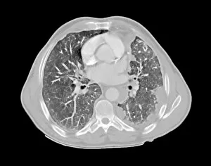
Background imagePulmonary Collection: Mesothelioma lung cancer, CT scan

Mesothelioma lung cancer, CT scan
Mesothelioma lung cancer. Computed tomography (CT) scan of the lungs of a 69 year old patient with mesothelioma, a type of lung cancer

Background imagePulmonary Collection: Respiratory system, artwork C015 / 6453

Respiratory system, artwork C015 / 6453
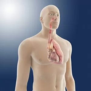
Respiratory system. Computer artwork showing the organs of the human respiratory system. At top is the larynx (voicebox), which leads onto the trachea (windpipe, white tube vertical)

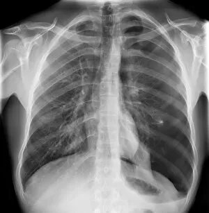
Background imagePulmonary Collection: Pneumothorax, X-ray C017 / 7966

Pneumothorax, X-ray C017 / 7966
Pneumothorax. X-ray of the chest of a 22 year old male patient with a pneumothorax, or collapsed lung. The left lung (right on X-ray) has collapsed due to a build-up of air (solid grey)

Background imagePulmonary Collection: Lung cancer, X-ray C017 / 7964

Lung cancer, X-ray C017 / 7964
Lung cancer. X-ray if the chest of a patient with malignant (cancerous) tumours in middle of the left lung (seen at right). The main cause of lung cancer is cigarette smoke

Background imagePulmonary Collection: Removed lung, X-ray C017 / 7823

Removed lung, X-ray C017 / 7823
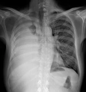
Removed lung. X-ray of the chest of a patient that has had their right lung (at left) removed

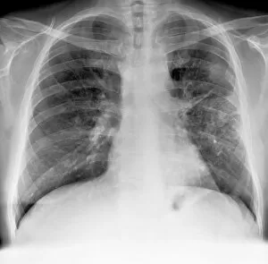
Background imagePulmonary Collection: Lung disease, X-ray C017 / 7817

Lung disease, X-ray C017 / 7817
Lung disease. X-ray of the chest of a patient with chronic obstructive pulmonary disease (COPD). This is a collection of diseases that includes emphysema and chronic bronchitis
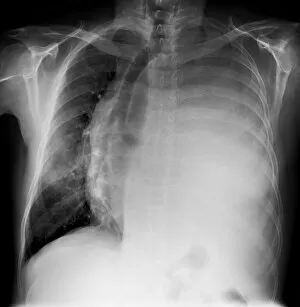
Background imagePulmonary Collection: Pleural effusion, X-ray C017 / 7811

Pleural effusion, X-ray C017 / 7811
Pleural effusion. X-ray of the chest of a patient with pleural effusion of the left lung (seen on right). Pleural effusion is the accumulation of fluid in the spaces around the lungs

Background imagePulmonary Collection: Pleural effusion, X-ray C017 / 7806

Pleural effusion, X-ray C017 / 7806
Pleural effusion. X-ray of the chest of a patient with bilateral pleural effusion. Pleural effusion is the accumulation of fluid in the spaces around the lungs

Background imagePulmonary Collection: Mesothelioma lung cancer, X-ray C017 / 7776

Mesothelioma lung cancer, X-ray C017 / 7776
Mesothelioma lung cancer, X-ray of the chest of a 84 year old male patient with advanced mesothelioma, a type of lung cancer

Background imagePulmonary Collection: Pleural effusion, X-ray C017 / 7753

Pleural effusion, X-ray C017 / 7753
Pleural effusion. X-ray of the chest of a 60 year old male patient with pleural effusion of the right lung (seen on left)

Background imagePulmonary Collection: Lung cancer, X-ray C016 / 4435

Lung cancer, X-ray C016 / 4435
Lung cancer. Coloured chest X-ray of a 64 year old male patient with lung cancer. The tumour is seen as the red mass in the right lung (bright area at left)

Background imagePulmonary Collection: Lung cancer, X-ray C016 / 4432

Lung cancer, X-ray C016 / 4432
Lung cancer. Coloured chest X-ray of a 64 year old male patient with lung cancer. The tumour is seen as the orange mass in the right lung (dark area at left)

Background imagePulmonary Collection: Lung cancer, X-ray C016 / 4433

Lung cancer, X-ray C016 / 4433
Lung cancer. Coloured chest X-ray of a 64 year old male patient with lung cancer. The tumour is seen as the orange mass in the right lung (bright area at left)

Background imagePulmonary Collection: Heart and lung anatomy, artwork C016 / 2911

Heart and lung anatomy, artwork C016 / 2911
Heart and lung anatomy. Artwork of an oblique anterior (left-frontal) view of the rib-cage, heart and lungs in a male body. One lung is shown at right

Background imagePulmonary Collection: Heart and lung anatomy, artwork C016 / 2909

Heart and lung anatomy, artwork C016 / 2909
Heart and lung anatomy. Artwork of an anterior (frontal) view of the heart and lungs in a male body. One lung is shown at right. The bronchioles (airways) inside the lungs are shown at left

Background imagePulmonary Collection: Lung anatomy, artwork C016 / 2905

Lung anatomy, artwork C016 / 2905
Lung anatomy. Artwork of an anterior (frontal) view of the lungs in a male body. One lung is shown at right. The heart is not shown

Background imagePulmonary Collection: Heart and lung anatomy, artwork C016 / 2904

Heart and lung anatomy, artwork C016 / 2904
Heart and lung anatomy. Artwork of a posterior (from behind) view of the heart and lungs. The heart (mostly obscured) is a hollow muscle that pumps blood around the body and to the lungs

Background imagePulmonary Collection: Thermogene heat padding advert, 1909 C016 / 2554

Thermogene heat padding advert, 1909 C016 / 2554
Thermogene heat padding advert. This padding, applied to the chest, was described as a counter-irritant calorific cotton wadding impregnated with vegetable essences

Background imagePulmonary Collection: Paediatric examination C013 / 8824

Paediatric examination C013 / 8824
Paediatric examination. Doctor using a stethoscope to listen to a 3-year-old boys lungs. A stethoscope is used to listen to sounds within the body

Background imagePulmonary Collection: Heswall Tuberculosis Sanatorium, Liverpool

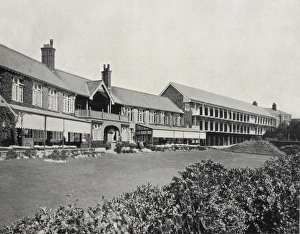
Heswall Tuberculosis Sanatorium, Liverpool
Heswall Sanatorium, jointly established in 1902 by Liverpool Parish, West Derby Union, and Toxteth Park Township for the treatment of children with phthisis - pulmonary tuberculosis

Background imagePulmonary Collection: Carpet beetle hair, SEM

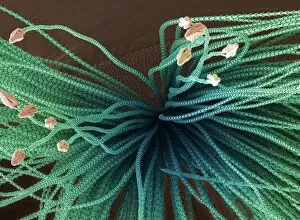
Carpet beetle hair, SEM
Carpet beetle hair. Coloured scanning electron micrograph (SEM) of the spear-headed hair (hastisetae) of a carpet beetle larva (family Dermestidae)

Background imagePulmonary Collection: Lungs

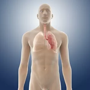
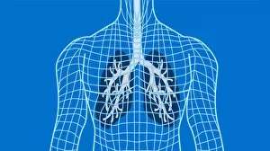
Lungs in a wireframe body, computer artwork. The windpipe (trachea, grey) runs downwards from the neck. It branches (upper centre) into two bronchi, one for each of the two lungs

Background imagePulmonary Collection: Lungs

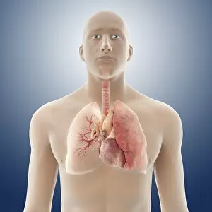
Lungs. Computer artwork of a naked woman with a healthy pair of lungs (red). The trachea (windpipe) is seen at the centre top of the chest. This splits into two bronchi, one for each lung

Background imagePulmonary Collection: Bronchioles

Bronchioles
Bronchiole network, composite image. The trachea (windpipe, not seen) branches into two bronchi, which enter the pair of lungs

Background imagePulmonary Collection: Lung, X-ray

Lung, X-ray
Lung. Coloured bronchography (X-ray) of a healthy human lung. A contrast medium has been added to show the network of airways (green) in the right lung

Background imagePulmonary Collection: Trachea epithelium, light micrograph

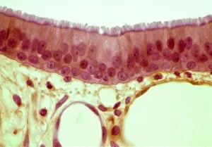
Trachea epithelium, light micrograph
Trachea epithelium. Light micrograph of a vertical section through the pseudostratified columnar epithelium from the trachea

Background imagePulmonary Collection: Lung bronchogram, coloured X-ray

Lung bronchogram, coloured X-ray
Healthy lung. Coloured bronchogram x-ray of a side (lateral) view of a healthy human lung. The vertebrae of the spinal column are seen at left

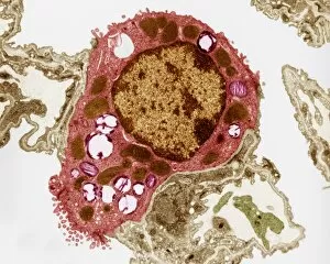
Background imagePulmonary Collection: Alveolar cell, TEM

Alveolar cell, TEM
Alveolar cell. Coloured transmission electron micrograph (TEM) of a section through a type II alveolar cell. This cell is found in the alveoli (air sacs) of the lungs

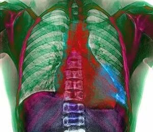
Background imagePulmonary Collection: Lungs and heart, X-ray

Lungs and heart, X-ray
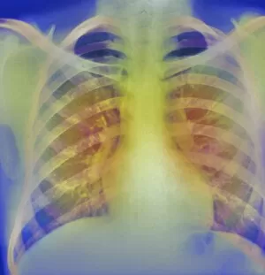
Lungs and heart, coloured chest X-ray. The lungs and heart are enclosed and protected by the ribs (horizontal bars) which meet at the breastbone, or sternum (down centre)

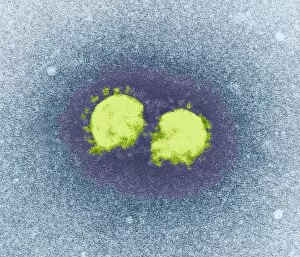
Background imagePulmonary Collection: SARS virus particles, TEM

SARS virus particles, TEM
SARS virus particles. Coloured transmission electron micrograph (TEM) of two SARS virus particles (green). Severe acute respiratory syndrome (SARS)

Background imagePulmonary Collection: Lung infection

Lung infection. Coloured chest X-ray of a patient with sickle cell disease (SCD), an inherited blood disorder. SCD has many symptoms including pulmonary complications

Background imagePulmonary Collection: Sarcoidosis of the lungs, X-ray

Sarcoidosis of the lungs, X-ray
Sarcoidosis. Frontal coloured chest X-ray of the lungs of a patient with pulmonary sarcoidosis. Affected lung tissue shows up as red patches

Background imagePulmonary Collection: Severe acute respiratory syndrome (SARS)

Severe acute respiratory syndrome (SARS). Conceptual image of the letters of SARS on a lung artwork. This represents the SARS virus infecting the lungs

Background imagePulmonary Collection: Lung scarring from tuberculosis, X-ray

Lung scarring from tuberculosis, X-ray
Tuberculosis lung scarring. Coloured chest X-ray showing scarring (green) of the lungs after a case of chronic tuberculosis (TB). The heart (red) is also seen

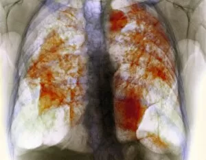
Background imagePulmonary Collection: Lung silicosis, X-ray

Lung silicosis, X-ray
Lung silicosis. Coloured X-ray of the lungs of a patient with silicosis, a type of pneumoconiosis. The orange masses in the lungs are areas of fibrous (scarred) tissue and inflammation

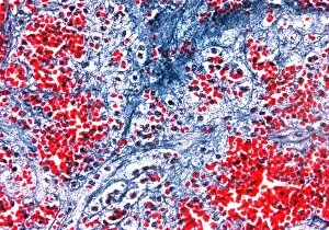
Background imagePulmonary Collection: Bleeding in emphysema

Bleeding in emphysema
Internal bleeding in lung tissue due to pulmonary emphysema, light micrograph. The bleeding is seen as numerous red blood cells (red)

Background imagePulmonary Collection: Emphysema of the lungs, CT scan

Emphysema of the lungs, CT scan
Emphysema. Coloured 3-D computed tomography (CT) scan of the lungs, showing advanced emphysema of the right lung (left). Emphysema is a disease in which the tiny lung air sacs (alveoli)

Background imagePulmonary Collection: Heart of gold

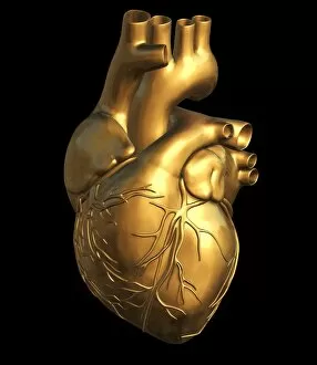
Heart of gold, computer artwork. The heart is a hollow sac (lower frame) of muscular tissue that pumps blood around the body. Small surface coronary blood vessels supply the heart muscle with blood

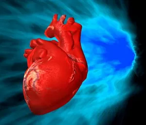
Background imagePulmonary Collection: Heart

Heart and glowing light, computer artwork. The heart (red) is a hollow sac of muscular tissue that pumps blood around the body

Background imagePulmonary Collection: Heart of glass

Heart of glass, computer artwork. The heart is a hollow muscle that pumps blood around the body. The vena cava vein (upper left) carries deoxygenated blood into the heart from the body

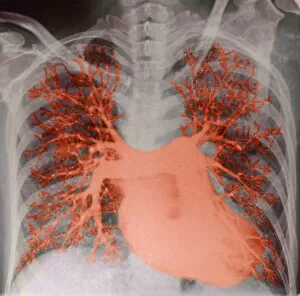
Background imagePulmonary Collection: Lung blood vessels, X-ray

Lung blood vessels, X-ray
Lung blood vessels. Coloured angiogram of the heart and pulmonary arteries. The heart is at lower right. The left and right pulmonary arteries branch from the top of the heart

Background imagePulmonary Collection: Heart and lungs

Heart and lungs. Historical artwork of the heart (pink) and lungs (red, left and right) seen from behind. Dissection hooks have been used to draw back the lung tissue

Background imagePulmonary Collection: Lung function test trace

Lung function test traces. Pen tracing the results from a lung function test onto graph paper. It is measuring the maximum rate at which air is expelled from the lungs

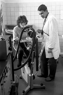
Background imagePulmonary Collection: Breathing research

Breathing research. Medical researcher observing a woman exercising as equipment is used to monitor her breathing. This is a functional diagnostics laboratory in Novosibirsk, Siberia, Russia

Background imagePulmonary Collection: Chronic bronchitis, X-ray

Chronic bronchitis, X-ray
Chronic bronchitis, coloured X-ray. This condition almost exclusively affects smokers. It is often associated with emphysema, which is known as COLD (chronic obstructive lung disease)

Background imagePulmonary Collection: Bacterial pneumonia infection

Bacterial pneumonia infection. Light micrograph of pneumonia bacteria (Klebsiella pneumoniae, rod- shaped, pink) among cells in a sample of pus from a pneumonia patient. K

Background imagePulmonary Collection: Childhood asthma

Childhood asthma. Artwork of a childs respiratory system showing the mucus (yellow) and narrowing of the airways (bronchioles) in paediatric asthma

Background imagePulmonary Collection: Deep vein thrombosis, artwork

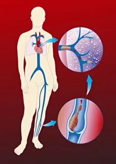
Deep vein thrombosis, artwork
Deep vein thrombosis. Computer artwork showing blood clots in a leg (brown lumps, bottom right), caused by deep vein thrombosis (DVT), moving to blood vessels in the lungs (blue, top right)


For sale as Licensed Images

Choose your image, Select your licence and Download the media

"Pulmonary: Unveiling the Intricacies of our Vital Breath" Discover the wonders of the pulmonary system through a captivating journey. Dive into a world where life-sustaining breaths take center stage, as we explore various aspects that define our respiratory health. Embark on this educational voyage with a detailed diagram showcasing the intricate structure of lungs and bronchial tubes. Marvel at their complexity, understanding how they facilitate oxygen exchange within our bodies. Witness medical advancements in pneumothorax treatment through X-ray imagery, offering hope to those affected by this condition. Delve into neck anatomy studies, unraveling the secrets hidden beneath our skin while appreciating 19th Century artwork depicting these discoveries. Uncover the devastating impact of tuberculosis through haunting X-ray images that reveal its presence within afflicted lungs. Gain insight into how interconnected systems work together harmoniously with an enlightening diagram illustrating the heart, lungs, and windpipe. Experience tension pneumothorax's urgency via gripping X-rays that depict its severity and prompt intervention requirements. Grasp the symbiotic relationship between heart and lungs; two vital organs intricately linked for optimal functioning. Explore cystic fibrosis' challenges faced by patients daily; understand their resilience amidst adversity. Learn about Konstantin Buteyko's groundbreaking contributions as a Soviet doctor revolutionizing respiratory therapy techniques. Marvel at abdominal arteries' inner workings revealed in an illuminating X-ray image (P206 / 0309), highlighting their significance in maintaining overall health. Discover Hantavirus' impact on pulmonary health – raising awareness about potential risks lurking in nature's realm. Reflect upon "HOCHE DIES AT WETZLAR, " reminding us of historical milestones shaping modern medicine's understanding diseases throughout time. Join us on this extraordinary expedition exploring every facet of "pulmonary. " Let curiosity guide you as we unravel mysteries surrounding respiration - an awe-inspiring process essential to our very existence.