Reproductive Collection (#7)

"Exploring the Intricate World of Reproduction: From Dinosaurs to Flowers and Beyond" In the ancient world

Background imageReproductive Collection: Zombie ant fungus life-cycle, artwork

Zombie ant fungus life-cycle, artwork
Zombie ant fungus life-cycle. Computer artwork showing the life cycle of the parasitoidal fungus Ophiocordyceps unilateralis

Background imageReproductive Collection: Follicle-stimulating hormone complex C016 / 2794

Follicle-stimulating hormone complex C016 / 2794
Follicle-stimulating hormone (FSH) complex with receptor, molecular model. FSH helps to regulate human sexual development and reproductive processes. In females, it acts on follicles in the ovaries

Background imageReproductive Collection: Follicle-stimulating hormone complex C016 / 2793

Follicle-stimulating hormone complex C016 / 2793
Follicle-stimulating hormone (FSH) complex with receptor, molecular model. FSH helps to regulate human sexual development and reproductive processes. In females, it acts on follicles in the ovaries
Background imageReproductive Collection: Mythical creature, 16th century C013 / 7648

Mythical creature, 16th century C013 / 7648
Mythical creature. 16th-century artwork of a mythical sucarath creature described as strange and dangerous beast. Its tail here is shown shielding its young, being carried on its back
Background imageReproductive Collection: Waved albatross courtship display C013 / 7508

Waved albatross courtship display C013 / 7508
Waved albatross courtship display. Pair of blue-waved albatross (Phoebastria irrorata) displaying during a courtship ritual

Background imageReproductive Collection: Chinstrap penguin courting display C013 / 7483

Chinstrap penguin courting display C013 / 7483
Chinstrap penguin courting display. Pair of chinstrap penguins (Pygoscelis antarcticus) displaying during a courtship ritual

Background imageReproductive Collection: Blue-footed booby and eggs C013 / 7473

Blue-footed booby and eggs C013 / 7473
Blue-footed booby and eggs. Close-up of a blue-footed booby (Sula nebouxii) with its eggs in a nest. Photographed on San Cristobal Island, Galapagos Islands

Background imageReproductive Collection: Blue-footed booby courtship display C013 / 7472

Blue-footed booby courtship display C013 / 7472
Blue-footed booby courtship display. Pair of blue-footed boobies (Sula nebouxii) displaying during a courtship ritual. Photographed on San Cristobal Island, Galapagos Islands

Background imageReproductive Collection: Ant symbiosis C013 / 7071

Ant symbiosis C013 / 7071
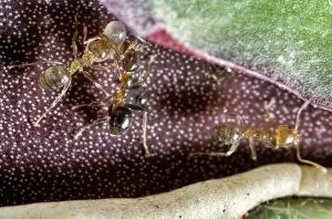
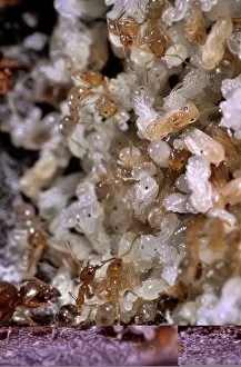
Ant symbiosis. Close-up of Philidris sp. ants and their eggs (white) inside the leaf of a Malayan urn vine (Dischidia major), with which they form a symbiotic relationship

Background imageReproductive Collection: Ant symbiosis C013 / 7073

Ant symbiosis C013 / 7073
Ant symbiosis. Close-up of Philidris sp. ants with their larvae (white) inside the leaf of a Malayan urn vine (Dischidia major), with which they form a symbiotic relationship

Background imageReproductive Collection: Ant symbiosis C013 / 7070

Ant symbiosis C013 / 7070
Ant symbiosis. Philidris sp. ants and their eggs (white) inside the leaf of a Malayan urn vine (Dischidia major), with which they form a symbiotic relationship

Background imageReproductive Collection: Trap-jaw ant carrying eggs C013 / 7056

Trap-jaw ant carrying eggs C013 / 7056
Trap-jaw ant carrying eggs. Close-up of a trap-jaw ant (Odontomachus bauri) using its large mandibles to carry eggs. This Central

Background imageReproductive Collection: Trap-jaw ant carrying eggs C013 / 7055

Trap-jaw ant carrying eggs C013 / 7055
Trap-jaw ant carrying eggs. Close-up of a trap-jaw ant (Odontomachus bauri) using its large mandibles to carry eggs. This Central

Background imageReproductive Collection: Verreauxs sifaka mother and baby

Verreauxs sifaka mother and baby
Verreauxs sifaka (Propithecus verreauxi) mother and baby on the ground. All lemurs are endemic to Madagascar, where they evolved in isolation from other primates after Madagascar became

Background imageReproductive Collection: Siamese Pig, 19th century C013 / 6233

Siamese Pig, 19th century C013 / 6233
Siamese Pig. 19th-century artwork of a sow and piglets of the Siamese Pig, a breed of the domesticated pig (Sus scrofa domestica)

Background imageReproductive Collection: Ryeland Sheep, 19th century C013 / 6231

Ryeland Sheep, 19th century C013 / 6231
Ryeland Sheep. 19th-century artwork of a ram, ewe and lamb of the Ryeland Sheep, a breed of the domesticated sheep (Ovis aries)

Background imageReproductive Collection: Soft-woolled Welsh sheep, 19th century C013 / 6229

Soft-woolled Welsh sheep, 19th century C013 / 6229
Soft-woolled Welsh sheep. 19th-century artwork of ewes and a lamb of the soft-woolled sheep from Wales, a breed of the domesticated sheep (Ovis aries)

Background imageReproductive Collection: Black-faced Heath Sheep, 19th century C013 / 6230

Black-faced Heath Sheep, 19th century C013 / 6230
Black-faced Heath Sheep. 19th-century artwork of a ewe and lambs of the Black-faced Heath Sheep, a breed of the domesticated sheep (Ovis aries)

Background imageReproductive Collection: Alderney Cattle, 19th century C013 / 6223

Alderney Cattle, 19th century C013 / 6223
Alderney Cattle. 19th-century artwork of a cow and calf of the Alderney Cattle, a breed of the domesticated cow (Bos taurus)

Background imageReproductive Collection: Sperm cell anatomy, artwork C013 / 4648

Sperm cell anatomy, artwork C013 / 4648
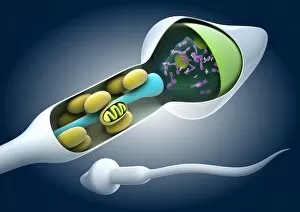
Sperm cell anatomy. Cutaway computer artwork showing the internal structure of a sperm cell (spermatozoon), the male sex cell

Background imageReproductive Collection: Fertilisation, artwork C013 / 4647

Fertilisation, artwork C013 / 4647
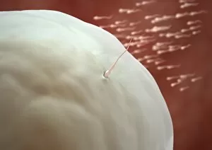
Fertilisation. Computer artwork of a sperm cell (spermatozoa) penetrating an egg (large)

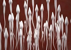
Background imageReproductive Collection: Sperm cells, artwork C013 / 4645

Sperm cells, artwork C013 / 4645
Sperm cells. Computer artwork of numerous sperm cells (spermatozoa), showing their torpedo-shaped heads, and long tails that they use for propulsion

Background imageReproductive Collection: Fertilisation, artwork C013 / 4646

Fertilisation, artwork C013 / 4646
Fertilisation. Computer artwork of numerous sperm cells (spermatozoa) swimming towards an egg (left)

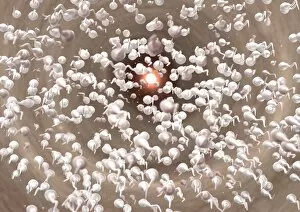
Background imageReproductive Collection: Fertilisation, artwork C013 / 4643

Fertilisation, artwork C013 / 4643
Fertilisation. Computer artwork of numerous sperm cells (spermatozoa) swimming towards an egg (bright) in the distance

Background imageReproductive Collection: Fertilisation, artwork C013 / 4644

Fertilisation, artwork C013 / 4644
Fertilisation. Computer artwork of numerous sperm cells (spermatozoa) swimming towards an egg (bright) in the distance

Background imageReproductive Collection: Alder tree pollen grain, SEM

Alder tree pollen grain, SEM
Alder tree pollen grain. Coloured scanning electron micrograph (SEM) of a pollen grain from an alder tree (Alnus glutinosa). A pollen grain contains a male sex cell from a flowering plant

Background imageReproductive Collection: Common poppy seeds (Papaver rhoeas) SEM

Common poppy seeds (Papaver rhoeas) SEM
Common poppy seeds (Papaver rhoeas), coloured scanning electron micrograph (SEM). Magnification x66 at 10cm wide

Background imageReproductive Collection: Chickens egg outer shell detail, SEM

Chickens egg outer shell detail, SEM
Chickens egg outer shell detail, coloured scanning electron micrograph (SEM). Hard-shelled eggs are produced by birds and some reptiles. The shell protects the growing embryo inside

Background imageReproductive Collection: Horsetail spores (Equisetum arvense) SEM

Horsetail spores (Equisetum arvense) SEM
Horsetail spores (Equisetum arvense) coloured scanning electron micrograph (SEM). The tendrils or elaters as they are known

Background imageReproductive Collection: Asiatic lily (Lilium asiatic) stigma, SEM

Asiatic lily (Lilium asiatic) stigma, SEM
Asiatic lily (Lilium asiatic) stigma detail, coloured scanning electron micrograph (SEM). Magnification of main image x15, inset image x332 at an image size of 10 cm

Background imageReproductive Collection: Lilium asiatic stigma detail, SEM

Lilium asiatic stigma detail, SEM
Asiatic lily (Lilium asiatic) stigma detail showing germinating pollen grains, coloured scanning electron micrograph (SEM). Magnification x465 at an image size of 10 cm

Background imageReproductive Collection: Flower bud detail, SEM

Flower bud detail, SEM
Detail of inside a flower bud, coloured scanning electron micrograph (SEM). The flower is a cape fuchsia (Phygelius aequalis). Magnification is x14 at an image of 10cm wide

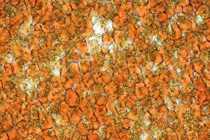
Background imageReproductive Collection: Jatropha curcas oil, light micrograph

Jatropha curcas oil, light micrograph
Jatropha curcas oil. Light micrograph of a section through a seed of the Barbados nut plant (Jatropha curcas), showing its oil content (orange). The granules stained yellow-green are protein

Background imageReproductive Collection: Jatropha curcas seeds

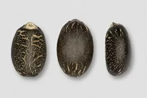
Jatropha curcas seeds. Oil extracted from the seeds of the Barbados nut plant (Jatropha curcas) is used as a biofuel, and can be further refined into biodiesel

Background imageReproductive Collection: Jatropha curcas seed

Jatropha curcas seed, transverse section. Oil extracted from the seeds of the Barbados nut plant (Jatropha curcas) is used as a biofuel, and can be further refined into biodiesel

Background imageReproductive Collection: Horsetail spore, SEM

Horsetail spore, SEM
Horsetail spore. Coloured scanning electron micrograph (SEM) of a spore of a horsetail (Equisetum arvense) plant. Spores (round) bear elaters (helical)

Background imageReproductive Collection: Broken egg shell, SEM

Broken egg shell, SEM
Broken egg shell, coloured scanning electron micrograph (SEM). Hard-shelled eggs are produced by birds and some reptiles. The shell protects the embryo inside

Background imageReproductive Collection: Egg shell membrane, SEM

Egg shell membrane, SEM
Egg shell membrane, coloured scanning electron micrograph (SEM). Broken egg shell, showing details of the membrane (across bottom) that is on the inside of the shell wall

Background imageReproductive Collection: Hounds tongue fruit

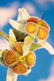
Hounds tongue fruits (Cynoglossum columnae). This plant is a member of the borage family. These are its unripe nutlets which usually occur in groups of four

Background imageReproductive Collection: Pomegranate (Punica granatum)

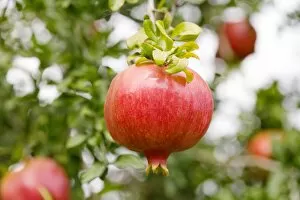
Pomegranate (Punica granatum)
Pomegranate fruit (Punica granatum) on its tree

Background imageReproductive Collection: Goosegrass fruit, SEM

Goosegrass fruit, SEM
Goosegrass fruit. Coloured scanning electron micrograph (SEM) of the hooked fruit of a goosegrass plant (Galium aparine). The hooks become lodged in the fur of passing animals

Background imageReproductive Collection: Foxglove seed, SEM

Foxglove seed, SEM
Foxglove seed. Coloured scanning electron micrograph (SEM) of the highly sculpted coat of a foxglove seed (Digitalis purpurea). Seed coat structure is often characteristic of a plant genera

Background imageReproductive Collection: Surface of strawberry fruit

Surface of strawberry fruit
Strawberry. Coloured scanning electron micrograph (SEM) of the surface of a strawberry. The flesh of the strawberry (Fragaria sp. ) is here coloured red

Background imageReproductive Collection: Coloured SEM of trichomes in orange peel

Coloured SEM of trichomes in orange peel
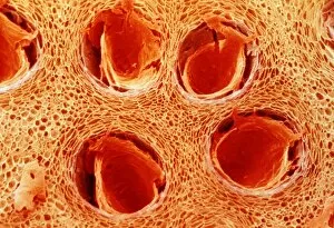
Orange peel. Coloured Scanning Electron Micrograph (SEM) of zest" cells in the peel of an orange, Citrus sp.. In this section the zest cells have been cut across and appear as the round

Background imageReproductive Collection: Willow herb seed pod, SEM

Willow herb seed pod, SEM
Willow herb seed pod. Coloured scanning electron micrograph (SEM) of willow herb (Epilobium montanum) seeds in their seed pod

Background imageReproductive Collection: Snapdragon seed, SEM

Snapdragon seed, SEM
Snapdragon (Antirrhinum majus) seed, coloured scanning electron micrograph (SEM). Small seeds like this one often have very roughly textured seed coats, or testas

Background imageReproductive Collection: Coloured SEM of trichomes in lemon peel

Coloured SEM of trichomes in lemon peel
Lemon peel. Coloured Scanning Electron Micrograph (SEM) of zest" cells in the peel of a lemon, Citrus limoni. In this section the zest cells have been cut across and appear as the round

Background imageReproductive Collection: Germinated seed, SEM

Germinated seed, SEM
Germinated seed. Image 4 of 4. Coloured scanning electron micrograph (SEM) of the final stage in the germination of a plant seed. The seed coat (testa) has been lost


For sale as Licensed Images

Choose your image, Select your licence and Download the media

"Exploring the Intricate World of Reproduction: From Dinosaurs to Flowers and Beyond" In the ancient world, even mighty Tyrannosaurus rex dinosaurs had their own unique ways of reproducing. Discovering fossilized evidence of their mating rituals offers a fascinating glimpse into the reproductive strategies employed by these prehistoric giants. Zooming in closer to modern times, microscopic wonders like lavender pollen grains under scanning electron microscopy (SEM) reveal intricate structures that play a crucial role in plant reproduction. Similarly, SEM images of geranium anthers and dahlia flower pollen showcase nature's attention to detail when it comes to ensuring successful pollination. Nature never ceases to amaze with its diverse range mechanisms. Take the cloudberry, for example - this Arctic fruit relies on cross-pollination facilitated by insects or wind for its continued existence. Moving beyond plants, we find ourselves exploring the realm of humans. The miracle of life unfolds within a pregnant woman's body as she nurtures new beginnings and prepares for motherhood. But reproduction is not limited to just larger organisms; even tiny creatures like honeybees have their own unique methods. SEM images capturing honeybee legs provide insight into how these industrious insects transport precious pollen from one flower to another, ensuring fertilization occurs. Venturing further into nature's hidden corners reveals captivating details about reproduction in unexpected places. Mushroom gills under SEM expose delicate structures designed for spore dispersal – a vital step in mushroom reproduction. Delving deeper still, tea flower stamens captured through SEM offer an up-close look at yet another facet of botanical fertility – showcasing intricate arrangements meant to attract pollinators and ensure successful seed production. Flowers continue enchanting us with their beauty as well as their reproductive prowess. Delphinium flowers boast vibrant colors while employing various strategies such as self-pollination or attracting specific pollinators like bees or butterflies.