Reproductive Collection (#9)

"Exploring the Intricate World of Reproduction: From Dinosaurs to Flowers and Beyond" In the ancient world

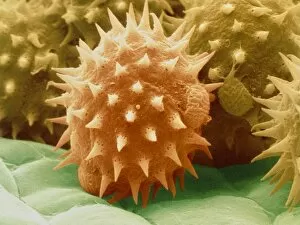
Background imageReproductive Collection: SEM of daisy pollen B786 / 0524

SEM of daisy pollen B786 / 0524
Daisy pollen. Coloured scanning electron micrograph (SEM) of pollen grains of a common Daisy (Bellis perennis). The outer wall (exine) has many small spiky projections

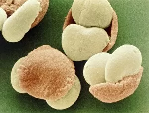
Background imageReproductive Collection: Pollen grains of Scots Pine tree

Pollen grains of Scots Pine tree
Pine pollen. Coloured scanning electron micrograph (SEM) of Scots Pine (Pinus sylvestris) pollen grains. Each pollen grain consists of a base (coloured brown) with two air-filled

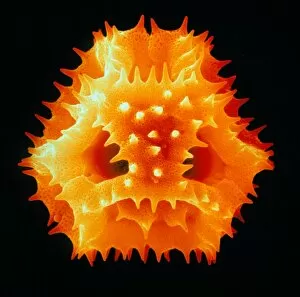
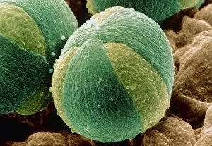
Background imageReproductive Collection: Pollen grain of a German chamomile flower

Pollen grain of a German chamomile flower
Chamomile pollen. Coloured scanning electron micrograph (SEM) of a single tricolporate pollen grain from a German chamomile flower, Matricaria recutita

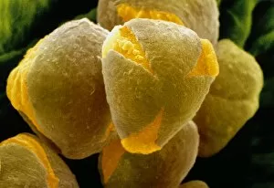
Background imageReproductive Collection: Pollengrainsfromthe flowering currant

Pollengrainsfromthe flowering currant
Currant pollen. Coloured scanning electron micrograph (SEM) of pollen grains from the flowering currant (Ribes sanguineum). This is a popular garden plant, and flowers appear in Spring

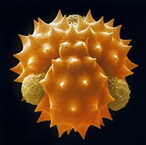
Background imageReproductive Collection: Pollen of the dandelion

Pollen of the dandelion
Dandelion pollen. Coloured scanning electron micrograph (SEM) of a pollen grain from the dandelion, Taraxacum officinale. The pollen grain is spined and finely-sculpted in shape

Background imageReproductive Collection: Tricolporate pollen of rose flower

Tricolporate pollen of rose flower
Rose pollen. Coloured scanning electron micrograph (SEM) of tricolporate pollen grains of a Garden Rose, Rosa sp. Pollen grains are the male sexual spores that are discharged from the male part of

Background imageReproductive Collection: Coloured SEM of sunflower pollen grains

Coloured SEM of sunflower pollen grains
Pollen grains. Coloured Scanning Electron Micro- graph of pollen grains of the sunflower, Helianthus annuus. Pollen grain size, shape and surface texture differ from one plant species to another

Background imageReproductive Collection: Timothy grass pollen grains

Timothy grass pollen grains
Timothy grass pollen. Coloured scanning electron micrograph (SEM) of a pollen grain from Timothy grass, Phelum pratense, also known as cat s-tail

Background imageReproductive Collection: Pollen grain of Sycamore tree

Pollen grain of Sycamore tree
Pollen grain. Coloured scanning electron micrograph (SEM) of pollen grains of the Sycamore tree, Acer pseudoplatanus. They are slightly triangularly shaped with coat ornamentation

Background imageReproductive Collection: Winged pollen grains from Lebanon cedar

Winged pollen grains from Lebanon cedar
Light micrograph of winged pollen grains from a cedar of Lebanon tree, Cedrus libani. Magnification: x400 at 35mm size

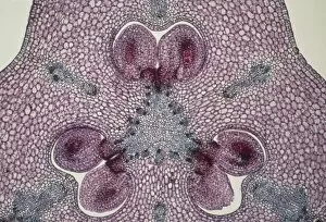
Background imageReproductive Collection: Section through a lily ovary with ovules

Section through a lily ovary with ovules
Light micrograph of a transverse section through the ovary of a lily, Lilium sp.. Two ovules (female gametes) are visible within each of the three hollow chambers of the ovary

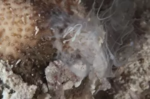
Background imageReproductive Collection: Sea Cucumber (Bohadschia marmorata) adult, ejecting sperm, Waigeo Island, Raja Ampat, West Papua

Sea Cucumber (Bohadschia marmorata) adult, ejecting sperm, Waigeo Island, Raja Ampat, West Papua, New Guinea, Indonesia

Background imageReproductive Collection: Liver flukes, transverse sections

Liver flukes, transverse sections
Liver flukes. Light micrographs of transverse sections through the male (top) and female (bottom) body areas of an adult liver fluke (Fasciola hepatica)

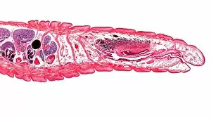
Background imageReproductive Collection: Earthworm, longitudinal section

Earthworm, longitudinal section
Earthworm. Light micrograph of a longitudinal section through the body of a round segmented earthworm (Lumbricus terrestris), showing the first 14 anterior segments

Background imageReproductive Collection: Liver fluke, transverse section

Liver fluke, transverse section
Liver fluke. Light micrograph of a transverse section through the anterior half of a liver fluke (Fasciola hepatica), a hermaphrodite tremotode worm and endoparasite that infects sheep

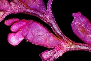
Background imageReproductive Collection: Campanularia hydrozoan, light micrograph

Campanularia hydrozoan, light micrograph
Campanularia hydrozoan. Polarised light micrograph of a hydroid colony of Campanularia polyps. The branches of the stem give rise to either feeding polyps (hydranths)

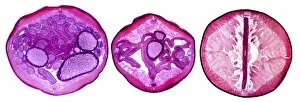
Background imageReproductive Collection: Nematode worm, transverse sections

Nematode worm, transverse sections
Nematode worm. Light micrograph of three transverse sections through the body of a female round unsegmented worm (Ascaris lumbricoides)

Background imageReproductive Collection: Hydroid (Campanularia flexuosa), LM

Hydroid (Campanularia flexuosa), LM
Hydroid (Campanularia flexuosa). Light micrograph (LM) of a colonial hydroid, showing gonozooid (reproducing) and gastrozooid (feeding) polyps, attached to a central stolon (stalk)

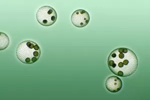
Background imageReproductive Collection: Green algae colonies

Green algae colonies. Light micrograph of green algae colonies (Volvox sp.). These colonies are made up of approximately 500 individual flagellate cells, arranged in a glycoprotein filled sphere
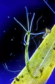
Background imageReproductive Collection: Freshwater Hydra, light micrograph

Freshwater Hydra, light micrograph
Freshwater Hydra. Dark-field light micrograph of a freshwater Hydra on a leaf of Canadian pondweed (Elodea canadensis). The Hydra is undergoing asexual reproduction by budding off a new individual

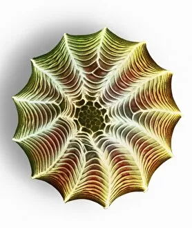
Background imageReproductive Collection: Cubic close-packed crystal structure

Cubic close-packed crystal structure
Haemoglobin molecule. Computer artwork showing the structure of a haemoglobin molecule. Haemoglobin is a metalloprotein that transports oxygen around the body in red blood cells

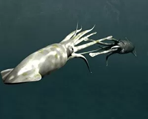
Background imageReproductive Collection: Squid mating

Squid mating. Computer artwork of two squid using their arms and tentacles to mate. During mating, the male places his sperm in a pouch on the female body

Background imageReproductive Collection: Pregnant female scorpion

Pregnant female scorpion. Scorpions give birth to live young after a gestation period ranging from a few months to over a year

Background imageReproductive Collection: Small white butterfly egg, SEM

Small white butterfly egg, SEM
Small white butterfly egg. Coloured scanning electron micrograph (SEM) of an egg of the small white butterfly (Pieris rapae)

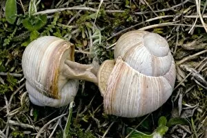
Background imageReproductive Collection: Roman snails mating

Roman snails mating
Roman snails (Helix pomatia) mating. Photographed in Sweden

Background imageReproductive Collection: Thick-billed weaver nest

Thick-billed weaver nest. This nest was built by the thick-billed weaver (Amblyospiza albifrons). This bird breeds in reeds

Background imageReproductive Collection: American robin nest

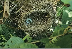
American robin nest. Empty nest with the broken shell of an egg from an American robin (Turdus migratorius). The female builds the nest of grass and twigs, normally lays 2-3 eggs

Background imageReproductive Collection: African black oystercatcher chick

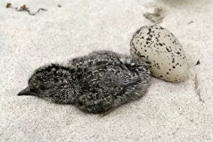
African black oystercatcher chick (Haematopus moquini) and an egg. The female lays a clutch of 1 to 2 eggs, and both male and female incubate the eggs for around 35 days

Background imageReproductive Collection: American robin chicks

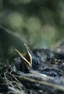
American robin chicks (Turdus migratorius) peering out of the nest. These birds are common throughout North America, living in woodland as well as in more built-up areas

Background imageReproductive Collection: Tadpole metamorphosis

Tadpole metamorphosis. Tadpole of the common frog (Rana temporaria). This tadpole is at the four leg stage (twelve weeks)

Background imageReproductive Collection: Common toads mating

Common toads mating (Bufo bufo). The male has climbed onto the females back. He will grasp the female for several days until she is ready to ovulate

Background imageReproductive Collection: Whale song

Whale song. Waveform traces of a short burst of the sounds made by a humpback whale (Megaptera novaeangliae). These sounds are produced by moving air back and forth through body passages

Background imageReproductive Collection: Southern elephant seals

Southern elephant seals
Southern elephant seal males (Mirounga leonina) sparring. Elephant seals are named for their snout or proboscis, which becomes enlarged during the breeding season

Background imageReproductive Collection: Bean aphid

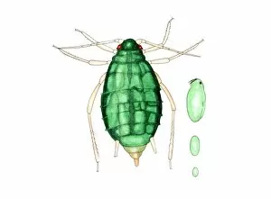
Bean aphid (Aphis fabae), artwork. This is a wingless female adult. It is able to reproduce parthenogentically (asexual reproduction), giving birth to pregnant offspring

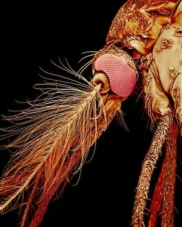
Background imageReproductive Collection: Male mosquito, SEM

Male mosquito, SEM
Male mosquito. Coloured scanning electron micrograph (SEM) of the head of an unidentified male mosquito (family Culicidae)

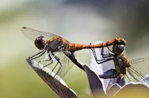
Background imageReproductive Collection: Common darter dragonflies

Common darter dragonflies (Sympetrum striolatum) mating on a flower. During mating the male dragonfly grasps the females head to pull her into the wheel position in order to copulate

Background imageReproductive Collection: Fly glandular organ, SEM

Fly glandular organ, SEM
Fly glandular organ. Coloured scanning electron micrograph (SEM) of a glandular organ on a March fly (family Bibionidae). This organ is found on the rear of the fly

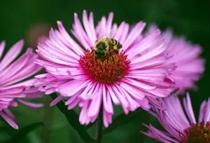
Background imageReproductive Collection: Honey bee on New England aster flower

Honey bee on New England aster flower
Honey bee (Apis melifera) on New England aster flower (Symphyotrichum novaeangliae)

Background imageReproductive Collection: Flightless dung beetle with dung ball

Flightless dung beetle with dung ball
Flightless dung beetle (Scarabaeus rugosus) on top of a dung ball. The dung beetles that roll dung into balls, known as rollers

Background imageReproductive Collection: Bee foot, SEM

Bee foot, SEM
Bee foot. Coloured scanning electron micrograph (SEM) of the tip of a leg from a bee (superfamily Apoidea). The end of an insect leg consists of the final segment, which is called the tarsus

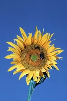
Background imageReproductive Collection: Bumblebees pollinating a sunflower

Bumblebees pollinating a sunflower
Bumblebees (Bombus sp.) pollinating a sunflower. As the bee feeds on nectar in the flower, pollen from the anthers become attached to it. Pollen grains are the male sex cells of a flowering plant

Background imageReproductive Collection: Blue-tailed damselfly

Blue-tailed damselfly
Blue-tailed damselflies (Ischnura elegans) mating on a plant stem. During mating the male damselfly grasps the females head to pull her into the wheel position in order to copulate

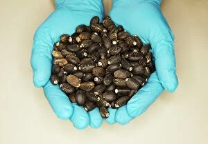
Background imageReproductive Collection: Biofuel research

Biofuel research. Gloved hands holding Jatropha curcas seeds. Oil extracted from the seeds of Jatropha plants is used as a biofuel, and can be further refined into biodiesel

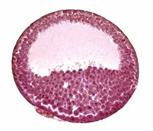
Background imageReproductive Collection: Frog embryo, light micrograph

Frog embryo, light micrograph
Frog embryo. Light micrograph of a frog embryo at the blastula stage. This stage in the embryos development is produced by the cleavage (cell division) of a fertilised ovum (egg cell)

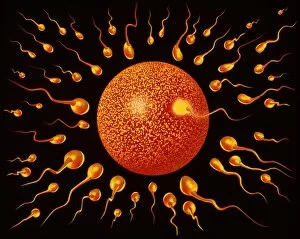
Background imageReproductive Collection: Sperm fertilising egg

Sperm fertilising egg

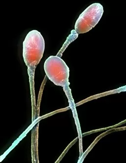
Background imageReproductive Collection: Human sperm

Human sperm

Background imageReproductive Collection: Female body anatomy

Female body anatomy, artwork

Background imageReproductive Collection: Male and female anatomy

Male and female anatomy. Historical artwork of male (left) and female (right) bodies showing their internal organs and circulatory systems


For sale as Licensed Images

Choose your image, Select your licence and Download the media

"Exploring the Intricate World of Reproduction: From Dinosaurs to Flowers and Beyond" In the ancient world, even mighty Tyrannosaurus rex dinosaurs had their own unique ways of reproducing. Discovering fossilized evidence of their mating rituals offers a fascinating glimpse into the reproductive strategies employed by these prehistoric giants. Zooming in closer to modern times, microscopic wonders like lavender pollen grains under scanning electron microscopy (SEM) reveal intricate structures that play a crucial role in plant reproduction. Similarly, SEM images of geranium anthers and dahlia flower pollen showcase nature's attention to detail when it comes to ensuring successful pollination. Nature never ceases to amaze with its diverse range mechanisms. Take the cloudberry, for example - this Arctic fruit relies on cross-pollination facilitated by insects or wind for its continued existence. Moving beyond plants, we find ourselves exploring the realm of humans. The miracle of life unfolds within a pregnant woman's body as she nurtures new beginnings and prepares for motherhood. But reproduction is not limited to just larger organisms; even tiny creatures like honeybees have their own unique methods. SEM images capturing honeybee legs provide insight into how these industrious insects transport precious pollen from one flower to another, ensuring fertilization occurs. Venturing further into nature's hidden corners reveals captivating details about reproduction in unexpected places. Mushroom gills under SEM expose delicate structures designed for spore dispersal – a vital step in mushroom reproduction. Delving deeper still, tea flower stamens captured through SEM offer an up-close look at yet another facet of botanical fertility – showcasing intricate arrangements meant to attract pollinators and ensure successful seed production. Flowers continue enchanting us with their beauty as well as their reproductive prowess. Delphinium flowers boast vibrant colors while employing various strategies such as self-pollination or attracting specific pollinators like bees or butterflies.