Research Collection (#93)

"Unveiling the Secrets

Background imageResearch Collection: Myoglobin molecule F006 / 9417

Myoglobin molecule F006 / 9417
Myoglobin, molecular model. Myoglobin is a protein found in muscle tissue, where it binds to and stores oxygen to be used during strenuous exercise

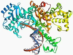
Background imageResearch Collection: Endonuclease and DNA, molecular model F006 / 9413

Endonuclease and DNA, molecular model F006 / 9413
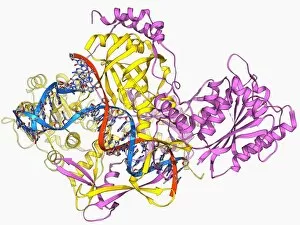
Endonuclease and DNA. Molecular model of an endonuclease restriction enzyme (yellow) bound to a molecule of DNA (deoxyribonucleic acid)

Background imageResearch Collection: Lactate dehydrogenase enzyme molecule F006 / 9411

Lactate dehydrogenase enzyme molecule F006 / 9411
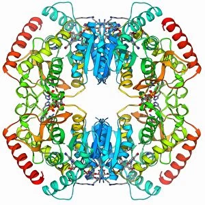
Lactate dehydrogenase enzyme, molecular model. This enzyme converts pyruvate to lactate in the final steps of glycolysis, and lactate to pyruvate during the lactic acid cycle

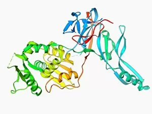
Background imageResearch Collection: Escherichia coli heat-labile enterotoxin F006 / 9410

Escherichia coli heat-labile enterotoxin F006 / 9410
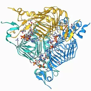
Escherichia coli heat-labile enterotoxin, molecular model. This is one of several proteins produced by pathogenic E. coli bacteria in the intestines

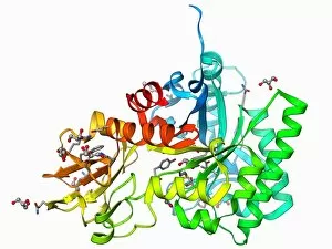
Background imageResearch Collection: Lipase molecule F006 / 9409

Lipase molecule F006 / 9409
Lipase molecule, molecular model. Lipase is an enzyme that breaks lipids (fats) into fatty acids and glycerol. Human pancreatic lipase is the main enzyme responsible for breaking down fat in

Background imageResearch Collection: Ubiquitin ligase molecule F006 / 9407

Ubiquitin ligase molecule F006 / 9407
Ubiquitin ligase, molecular model. This enzyme tags proteins for degradation by attaching a ubiquitin molecule to them

Background imageResearch Collection: Gene activator protein F006 / 9406

Gene activator protein F006 / 9406
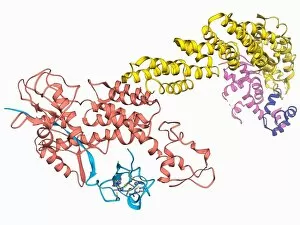
Gene activator protein. Molecular model of catabolite gene activator protein (CAP, yellow) complexed with deoxyribonucleic acid (DNA, red and blue) and RNA polymerase (green and pink)

Background imageResearch Collection: Microglobulin protein, molecular model F006 / 9408

Microglobulin protein, molecular model F006 / 9408
Microglobulin protein. Molecular model of the protein beta-2 microglobulin. This protein, with a relatively small molecular mass, is part of the immunoglobulin protein family

Background imageResearch Collection: Antibiotic resistance enzyme molecule F006 / 9405

Antibiotic resistance enzyme molecule F006 / 9405
Antibiotic resistance enzyme. Molecular model of an aminoglycoside phosphotransferase enzyme. This enzyme confers resistance to aminoglycoside antibiotics

Background imageResearch Collection: Cell adhesion protein molecule F006 / 9404

Cell adhesion protein molecule F006 / 9404
Cell adhesion protein. Molecular model of the ectodomain of the cell adhesion protein c-cadherin. This transmembrane protein facilitates adhesion between cells in solid tissues

Background imageResearch Collection: Cytochrome complex molecule F006 / 9401

Cytochrome complex molecule F006 / 9401
Cytochrome complex. Molecular model of cytochrome BC1 complex with bound cytochrome C. Cytochrome molecules perform oxidation and reduction reactions for electron transport

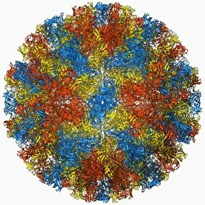
Background imageResearch Collection: Dengue virus capsid, molecular model F006 / 9392

Dengue virus capsid, molecular model F006 / 9392
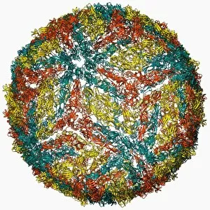
Dengue virus capsid, molecular model. This virus, transmitted by mosquito bites, causes the tropical disease dengue fever in humans

Background imageResearch Collection: Galactoside acetyltransferase molecule F006 / 9400

Galactoside acetyltransferase molecule F006 / 9400
Galactoside acetyltransferase, molecular model. This enzyme from the bacterium Escherichia coli is involved in the lac operon

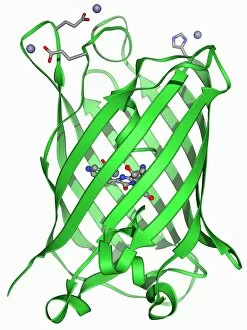
Background imageResearch Collection: Green fluorescent protein molecule F006 / 9402

Green fluorescent protein molecule F006 / 9402
Green fluorescent protein (GFP), molecular model. The molecule has a cylindrical structure formed from beta sheets (ribbons). GFP is found in the Pacific jellyfish Aequorea victoria

Background imageResearch Collection: Outer membrane receptor protein molecule F006 / 9398

Outer membrane receptor protein molecule F006 / 9398
Outer membrane receptor protein. Molecular model of FecA an outer membrane receptor protein

Background imageResearch Collection: Antibiotic resistance enzyme molecule F006 / 9399

Antibiotic resistance enzyme molecule F006 / 9399
Antibiotic resistance enzyme. Molecular model of kanamycin nucleotidyltransferase (KNTase). This bacterial enzyme is responsible for resistance to a number of antibiotics

Background imageResearch Collection: Enzyme catalysing DNA recombination F006 / 9395

Enzyme catalysing DNA recombination F006 / 9395
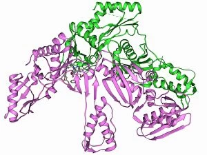
Enzyme catalysing DNA recombination. Molecular model of the enzyme CRE (cyclization recombination) recombinase (green and purple) mediating the recombination of strands of DNA (deoxyribonucleic acid)

Background imageResearch Collection: DNA polymerase Klenow fragment F006 / 9397

DNA polymerase Klenow fragment F006 / 9397
Klenow fragment of DNA polymerase I. Molecule model of the Klenow, or large, fragment from DNA polymerase I complexed with DNA (deoxyribonucleic acid, red and blue)

Background imageResearch Collection: Anthrax oedema factor molecule F006 / 9393

Anthrax oedema factor molecule F006 / 9393
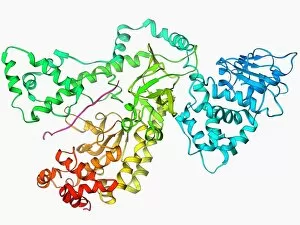
Anthrax oedema factor. Molecular model of oedema factor (EF) from the anthrax bacterium Bacillus antracis complexed with a calmodulin protein molecule

Background imageResearch Collection: Fish antifreeze protein F006 / 9396

Fish antifreeze protein F006 / 9396
Fish antifreeze protein. Molecular model of a fish antifreeze protein (AFP) from the ocean pout (Zoarces americanus)

Background imageResearch Collection: Proteinase inhibitor molecule F006 / 9394

Proteinase inhibitor molecule F006 / 9394
Proteinase inhibitor. Molecular model of a proteinase inhibitor, or antitrypsin, molecule bound to a tryspin protease. The proteinase inhibitor is a type of serine protease inhibitor (serpin)

Background imageResearch Collection: Voltage-gated potassium channel F006 / 9391

Voltage-gated potassium channel F006 / 9391
Voltage-gated potassium channel. Molecular model of a voltage-gated potassium (Kv) ion channel complexed with the antigen-binding fragment (Fab) of a monoclonal antibody

Background imageResearch Collection: DNA polymerase IV with DNA F006 / 9390

DNA polymerase IV with DNA F006 / 9390
polymerase IV with DNA. Molecular model of DNA polymerase IV (beige) complexed with a molecule of DNA (deoxyribonucleic acid, red and blue)

Background imageResearch Collection: Coxsackie B3 virus particle F006 / 9381

Coxsackie B3 virus particle F006 / 9381
Coxsackie B3 virus. Computer model of the capsid of the Coxsackie B3 virus

Background imageResearch Collection: Integrin, molecular model F006 / 9388

Integrin, molecular model F006 / 9388
Integrin. Molecular model of the integrin protein alpha-v beta-3. This is a transmembrane protein that is found on platelets. It is composed of two subunits; integrin alpha-V and integrin beta 3

Background imageResearch Collection: VMA-1 derived endonuclease molecule F006 / 9389

VMA-1 derived endonuclease molecule F006 / 9389
VMA-1 derived endonuclease. Molecular model of the VMA-1 derived endonuclease restriction enzyme. Restriction enzymes, also known as restriction endonucleases

Background imageResearch Collection: Uroporphyrinogen III decarboxylase F006 / 9387

Uroporphyrinogen III decarboxylase F006 / 9387
Uroporphyrinogen III decarboxylase. Molecular model of the enzyme human uroporphyrinogen III decarboxylase (UROD). Mutations or deficiencies in this enzyme cause the disorder porphyria with a build

Background imageResearch Collection: HIV reverse transcription enzyme F006 / 9385

HIV reverse transcription enzyme F006 / 9385
HIV reverse transcription enzyme. Molecular model of the reverse transcriptase enzyme found in HIV (the human immunodeficiency virus) bound to the inhibitor nevirapine

Background imageResearch Collection: Anthrax lethal factor molecule F006 / 9384

Anthrax lethal factor molecule F006 / 9384
Anthrax lethal factor, molecular model. This enzyme is one of three protein components that form the anthrax toxin produced by the bacterium Bacillus anthracis

Background imageResearch Collection: Early endosome antigen 1 molecule F006 / 9386

Early endosome antigen 1 molecule F006 / 9386
Early endosome antigen 1 (EEA1), molecular model. This protein is involved in the fusion and sorting of endosomes (membrane-bound compartments used to transport molecules)

Background imageResearch Collection: Manganese catalase enzyme molecule F006 / 9383

Manganese catalase enzyme molecule F006 / 9383
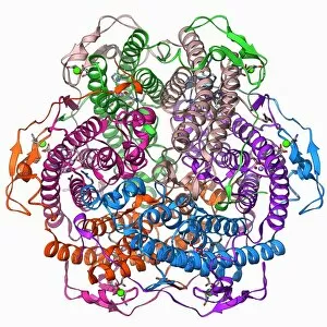
Manganese catalase. Molecular model of the enzyme manganese catalase from the bacterium Lactobacillus plantarum. This enzyme catalyses the break down of hydrogen peroxide to water and oxygen

Background imageResearch Collection: Photosystem I molecule F006 / 9380

Photosystem I molecule F006 / 9380
Photosystem I. Molecular model of the photosystem I complex from the cyanobacterium Synechococcus elongatus. Shown here are beta-carotene, alpha-chlorophyll and reaction centre subunits

Background imageResearch Collection: Repair protein and DNA, molecular model F006 / 9382

Repair protein and DNA, molecular model F006 / 9382
Repair protein and DNA. Molecular model of the Ku heterodimer (yellow and pink) bound to a strand of DNA (deoxyribonucleic acid, red and blue) as part of the repair process

Background imageResearch Collection: Norwalk virus capsid, molecular model F006 / 9372

Norwalk virus capsid, molecular model F006 / 9372
Norwalk virus capsid, molecular model. This norovirus, which causes a viral form of gastroenteritis, is transmitted from person-to-person or through contaminated food

Background imageResearch Collection: Anthrax lethal factor molecule F006 / 9379

Anthrax lethal factor molecule F006 / 9379
Anthrax lethal factor, molecular model. This enzyme is one of three protein components that form the anthrax toxin produced by the bacterium Bacillus anthracis

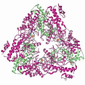
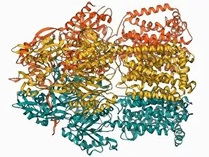
Background imageResearch Collection: Multidrug efflux pump molecule F006 / 9376

Multidrug efflux pump molecule F006 / 9376
Multidrug efflux pump. Molecular model of the multidrug efflux pump AcrB from the bacterium Escherichia coli. This protein pumps drugs, including antibiotics, out of the bacterial cell

Background imageResearch Collection: Calcium pumping ATPase muscle enzyme F006 / 9377

Calcium pumping ATPase muscle enzyme F006 / 9377
Calcium pumping ATPase enzyme. Molecular model of the ATPase enzyme that pumps calcium in and out of muscle cells and controls muscle contractions

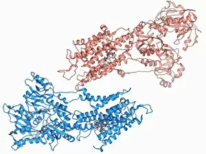
Background imageResearch Collection: Epidermal growth factor and receptor F006 / 9375

Epidermal growth factor and receptor F006 / 9375
Epidermal growth factor molecule. Molecular model of epidermal growth factor (EGF) bound to a receptor. EGF plays an important role in the regulation of cell growth, proliferation and differentiation

Background imageResearch Collection: Amyloid beta protein molecule F006 / 9378

Amyloid beta protein molecule F006 / 9378
Amyloid beta protein. Molecular model showing the structure of amyloid beta protein, the primary component of amyloid plaques found in the brains of Alzheimers patients

Background imageResearch Collection: Chitinase enzyme molecule F006 / 9374

Chitinase enzyme molecule F006 / 9374
Chitinase, molecular model. This enzyme catalyses the break down of glycosidic bonds in chitin, the main component of fungal cell walls and the exoskeletons of arthropods

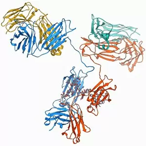
Background imageResearch Collection: Immunoglobulin G antibody molecule F006 / 9371

Immunoglobulin G antibody molecule F006 / 9371
Immunoglobulin G (IgG) antibody, molecular model. This is the most abundant immunoglobulin and is found in all body fluids

Background imageResearch Collection: Human interferon alpha molecule F006 / 9373

Human interferon alpha molecule F006 / 9373
Human interferon alpha, molecular model. Interferons are proteins produced by white blood cells as part of the immune response to invading pathogens, especially viruses

Background imageResearch Collection: DNA transcription, molecular model F006 / 9368

DNA transcription, molecular model F006 / 9368
DNA transcription. Molecular model of the enzyme RNA polymerase II synthesising a mRNA (messenger ribonucleic acid) strand from a DNA (deoxyribonucleic acid) template

Background imageResearch Collection: Hexokinase sugar enzyme F006 / 9370

Hexokinase sugar enzyme F006 / 9370
Hexokinase, molecular model. This enzyme promotes the conversion (phosphorylation) of glucose into glucose 6-phosphate. Cells then use the glucose 6-phosphate when they require energy

Background imageResearch Collection: Eye lens protein molecule F006 / 9367

Eye lens protein molecule F006 / 9367
Eye lens protein. Molecular model of delta-crystallin, a protein found in the lens of the eye. The regular arrangement of the protein in the lens is thought to be responsible for its transparency

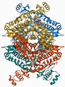
Background imageResearch Collection: Cholesterol producing enzyme and statin F006 / 9366

Cholesterol producing enzyme and statin F006 / 9366
Cholesterol producing enzyme and statin. Computer model showing the molecular structure of HMG-CoA reductase (HMGCR) in complex with Atorvastatin

Background imageResearch Collection: Interleukin-1 beta molecule F006 / 9369

Interleukin-1 beta molecule F006 / 9369
Interleukin-1 beta, molecular model. This cytokine immune protein is an important mediator of the inflammatory response

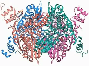
Background imageResearch Collection: Cholesterol producing enzyme molecule F006 / 9365

Cholesterol producing enzyme molecule F006 / 9365
Cholesterol producing enzyme. Molecular model of HMG-CoA reductase (HMGCR), the rate-controlling enzyme of the metabolic pathway that produces cholesterol in the body


For sale as Licensed Images

Choose your image, Select your licence and Download the media

"Unveiling the Secrets: Exploring the Vast Realm of Research" From deciphering the valorous tales behind British Military medals to unraveling the enigmatic patterns in a Rorschach Inkblot Test, research takes us on an intellectual odyssey. It delves into realms beyond our comprehension, like scrutinizing the cosmic microwave background through MAP and decoding intricate histological diagrams of a mammalian retina. Research is not confined to laboratories; it extends to observing nature's wonders firsthand. Jane Goodall, with her unwavering dedication as a British conservationist and zoologist, captured alongside a chimpanzee, epitomizes how research bridges gaps between species. The quest for knowledge transcends boundaries of time and space. Erwin Schrodinger's groundbreaking discoveries in quantum mechanics remind us that research propels humanity forward by challenging conventional wisdom. In particle physics, proton collisions such as C014 / 1797 or witnessing Higgs boson events like C013 / 6892 within ATLAS detectors demonstrate how researchers strive to unlock mysteries at subatomic levels. Pioneers like Marie Curie exemplify relentless pursuit despite adversities faced by women in science. Her revolutionary work with radioactivity continues to inspire generations. It also embarks on daring expeditions akin to HMS Beagle - Darwin's ship that sailed uncharted waters - pushing boundaries of exploration and understanding. Similarly, Apollo 17 astronauts left indelible footprints on the Moon while expanding human horizons beyond Earth's confines. These captivating glimpses into diverse facets highlight its transformative power. It fuels curiosity, challenges assumptions, and empowers us with knowledge that shapes our world. Whether investigating microscopic particles or exploring vast ecosystems – research illuminates paths towards progress and inspires future generations to embark upon their own quests for discovery.