Research Collection (#95)

"Unveiling the Secrets

Background imageResearch Collection: ITER Reaction vessel C018 / 3801

ITER Reaction vessel C018 / 3801
Artists concept view of the interior of the ITER reaction vessel. Nuclear fusion involves creating a plasma of superheated gas to temperatures of more than 200 million degrees C

Background imageResearch Collection: Ebola matrix protein molecule F006 / 9317

Ebola matrix protein molecule F006 / 9317
Ebola matrix protein. Molecular model of the Ebola virus matrix protein VP40. This membrane-associated protein is thought to be necessary for the assembly and budding of viral particles

Background imageResearch Collection: Insect antifreeze protein F006 / 9319

Insect antifreeze protein F006 / 9319
Insect antifreeze protein. Molecular model of an insect antifreeze protein (AFP) from the spruce budworm (Choristoneura fumiferana)

Background imageResearch Collection: Oxoguanine glycosylase complex F006 / 9318

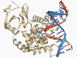
Oxoguanine glycosylase complex F006 / 9318
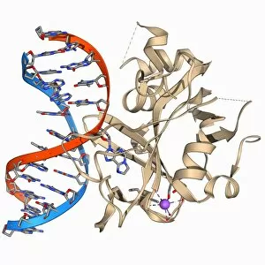
Oxoguanine glycosylase complex. Computer model showing an 8-Oxoguanine glycosylase (OGG1) molecule (beige) bound to a section of DNA (deoxyribonucleic acid, red and blue)

Background imageResearch Collection: Transcription repressor molecule F006 / 9316

Transcription repressor molecule F006 / 9316
Transcription repressor. Molecular model of the Tup 1 transcription repressor protein. Transcription repressors bind to specific sequences of DNA (deoxyribonucleic acid)

Background imageResearch Collection: Restriction enzyme and DNA F006 / 9315

Restriction enzyme and DNA F006 / 9315
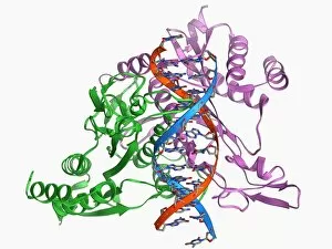
Restriction enzyme and DNA. Molecular model showing an EcoRI endonuclease enzyme (purple and green) bound to a DNA (deoxyribonucleic acid) molecule (red and blue)

Background imageResearch Collection: Green fluorescent protein molecule F006 / 9313

Green fluorescent protein molecule F006 / 9313
Green fluorescent protein (GFP), molecular model. The molecule has a cylindrical structure formed from beta sheets (ribbons). GFP is found in the Pacific jellyfish Aequorea victoria

Background imageResearch Collection: MoaC protein molecule F006 / 9312

MoaC protein molecule F006 / 9312
MoaC protein. Molecular model of the molybdenum cofactor biosynthesis protein MoaC. This enzyme is involved in carbon, nitrogen and sulphur metabolism

Background imageResearch Collection: Poliovirus particle F006 / 9306

Poliovirus particle F006 / 9306
Poliovirus particle, molecular model

Background imageResearch Collection: LAC repressor bound to DNA F006 / 9309

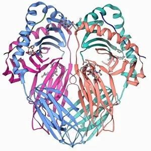
LAC repressor bound to DNA F006 / 9309
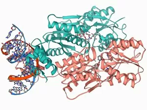
LAC repressor bound to DNA. Molecular model of a LAC (lactose) repressor molecule (pink and turquoise) interacting with bacterial DNA (deoxyribonucleic acid, red and blue)

Background imageResearch Collection: Epidermal growth factor molecule F006 / 9311

Epidermal growth factor molecule F006 / 9311
Epidermal growth factor (EGF), molecular model. EGF plays an important role in the regulation of cell growth, proliferation and differentiation

Background imageResearch Collection: Elongation factors Tu and Ts F006 / 9310

Elongation factors Tu and Ts F006 / 9310
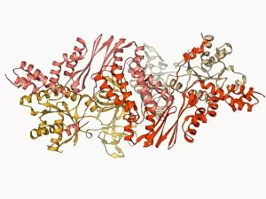
Elongation factors Tu and Ts, molecular model. These enzymes are involved in the elongation of polypeptide chains during translation

Background imageResearch Collection: Erythropoietin bound to receptors F006 / 9308

Erythropoietin bound to receptors F006 / 9308
Artwork of the human erythropoietin (EPO) hormone molecule (yellow) bound to receptors (pink). Erythropoietin regulates blood oxygen levels in the body

Background imageResearch Collection: Pyruvate dehydrogenase complex enzyme F006 / 9303

Pyruvate dehydrogenase complex enzyme F006 / 9303
Pyruvate dehydrogenase complex enzyme. Molecular model of dihydrolipoyl transacetylase one of the enzymes in the pyruvate dehydrogenase complex

Background imageResearch Collection: Oxoguanine glycosylase complex F006 / 9307

Oxoguanine glycosylase complex F006 / 9307
Oxoguanine glycosylase complex. Computer model showing an 8-Oxoguanine glycosylase (OGG1) molecule (beige) bound to a section of DNA (deoxyribonucleic acid, red and blue)

Background imageResearch Collection: Semliki forest virus capsid F006 / 9297

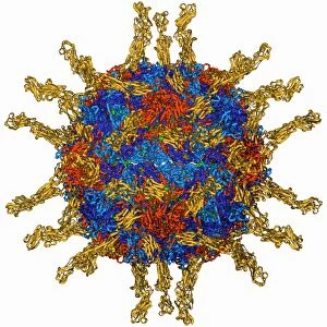
Semliki forest virus capsid F006 / 9297
Semliki forest virus capsid, molecular model. This virus, named for the forest in Uganda where it was identified, is spread by the bite of mosquitoes. It can infect both humans and animals

Background imageResearch Collection: ATPase molecule F006 / 9300

ATPase molecule F006 / 9300
ATPase molecule. Molecular model of the central stalk of an ATP synthase (ATPase) molecule from a cow. ATPase is an important enzyme that provides energy for cells through the synthesis of adenosine

Background imageResearch Collection: Cytochrome P450 and fluconazole F006 / 9302

Cytochrome P450 and fluconazole F006 / 9302
Cytochrome P450 and fluconazole. Molecular model of cytochrome P450 complexed with the anti-fungal compound fluconazole. This protein plays a crucial role in metabolism in animals (including humans)

Background imageResearch Collection: Bacteriorhodopsin protein F006 / 9299

Bacteriorhodopsin protein F006 / 9299
Bacteriorhodopsin protein. Molecular model showing the structure of bacteriorhodopsin (bR), a protein found in primitive micro-organisms known as Archaea. This protein acts as a proton pump

Background imageResearch Collection: Human serum albumin molecule F006 / 9301

Human serum albumin molecule F006 / 9301
Human serum albumin, molecular model. Albumin is the most abundant protein in human blood plasma. It is seen here complexed with stearic acid, a saturated fatty acid

Background imageResearch Collection: HP1 molecule C-terminal domain F006 / 9298

HP1 molecule C-terminal domain F006 / 9298
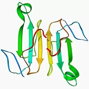
HP1 molecule C-terminal domain. Molecular model showing the structure of the C terminal (shadow chromo) domain of a heterochromatin protein 1 (HP1) molecule from a mouse

Background imageResearch Collection: Human poliovirus, molecular model F006 / 9289

Human poliovirus, molecular model F006 / 9289
Human poliovirus particle. Molecular model of the capsid of the human poliovirus. The capsid is a protein coat that encloses the viruss genetic information (genome), stored as RNA (ribonucleic acid)

Background imageResearch Collection: Green mamba venom molecule F006 / 9296

Green mamba venom molecule F006 / 9296
Green mamba venom. Molecular model of the alpha-dendrotoxin from green mamba (Dendroaspis angusticeps) venom

Background imageResearch Collection: MHC protein complexed with flu virus F006 / 9294

MHC protein complexed with flu virus F006 / 9294
MHC protein complexed with flu virus. Molecular model showing human class II MHC (major histocompatibility complex) protein HLA-DR1 complexed with an influenza (flu) virus peptide

Background imageResearch Collection: Hepatitis D virus ribozyme complex F006 / 9295

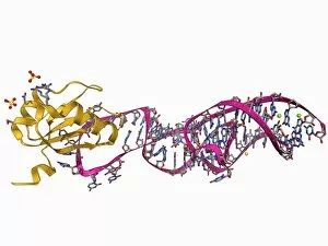
Hepatitis D virus ribozyme complex F006 / 9295
Hepatitis D virus ribozyme complex. Molecular model showing an RNA (ribonucleic acid) strand from an Hepatitis delta (Hepatitis D) virus genomic ribozyme, complexed with a ribonucleoprotein

Background imageResearch Collection: Heat shock protein 70 F006 / 9293

Heat shock protein 70 F006 / 9293
Heat shock protein 70. Molecular model of the 70 kDa heat shock protein (HSP) complexed with its substrate peptide. HSPs are a group of proteins whose levels increase when cells are exposed to raised

Background imageResearch Collection: Nucleotide exchange factor F006 / 9292

Nucleotide exchange factor F006 / 9292
Nucleotide exchange factor. Molecular model of the nucleotide exchange factor protein GrpE complexed with the chaperone protein DnaK

Background imageResearch Collection: Lumazine synthase molecule F006 / 9291

Lumazine synthase molecule F006 / 9291
Lumazine synthase molecule. Molecular model showing the structure of a lumazine synthase enzyme molecule from a Brucella abortus bacterium

Background imageResearch Collection: NAD-dependent DNA ligase molecule F006 / 9290

NAD-dependent DNA ligase molecule F006 / 9290
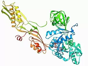
NAD dependent DNA ligase. Molecular model of NAD(+)-dependent DNA ligase. DNA ligase is an enzyme that binds two strands of DNA (deoxyribonucleic acid) together

Background imageResearch Collection: Human catalsae, molecular model F006 / 9288

Human catalsae, molecular model F006 / 9288
Human catalase, molecular model. This enzyme catalyses the break down of hydrogen peroxide to water and oxygen. Hydrogen peroxide is a highly toxic byproduct of a number of normal cellular processes

Background imageResearch Collection: Beta-carbonic anhydrase molecule F006 / 9286

Beta-carbonic anhydrase molecule F006 / 9286
Beta-carbonic anhydrase, molecular model. This enzyme is from the red algae Porphyridium purpureum. It catalyses the reversible hydration of carbon dioxide

Background imageResearch Collection: Ribonuclease bound to inhibitor F006 / 9287

Ribonuclease bound to inhibitor F006 / 9287
Ribonuclease bound to inhibitor, molecular model. Ribonuclease (RNase) is a type of nuclease that catalyses the degradation of RNA (ribonucleic acid)

Background imageResearch Collection: Elongation factor G F006 / 9284

Elongation factor G F006 / 9284
Elongation factor G. Molecular model of elongation factor G (EF-G) complexed with GDP (guanosine diphosphate). This enzyme is involved in the elongation of polypeptide chains during translation

Background imageResearch Collection: DNA Holliday junction, molecular model F006 / 9285

DNA Holliday junction, molecular model F006 / 9285
DNA Holliday junction. Molecular model of a Holliday junction (centre) between homologous strands of DNA (deoxyribonucleic acid)

Background imageResearch Collection: Coagulation factor complex molecule F006 / 9283

Coagulation factor complex molecule F006 / 9283
Coagulation factor complex molecule. Molecular model showing a blood clotting factor VIIa-tissue factor complex molecule. Tissue factor, also known as factor III

Background imageResearch Collection: Yeast DNA recognition, molecular model F006 / 9282

Yeast DNA recognition, molecular model F006 / 9282
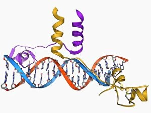
Yeast DNA recognition. Computer model showing a GAL4 transcription activator protein bound to a yeast DNA (deoxyribonucleic acid) molecule (red and blue)

Background imageResearch Collection: Creatine kinase molecule F006 / 9276

Creatine kinase molecule F006 / 9276
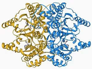
Creatine kinase molecule. This enzyme catalyses the phosphorylation of creatine to creatine phosphate. It plays an important role in energy metabolism in cells with high or fluctuating energy

Background imageResearch Collection: Human transferrin receptor molecule F006 / 9280

Human transferrin receptor molecule F006 / 9280
Human transferrin receptor, molecular model. This molecule is found on the surface of a cell. It binds transferrins, iron-binding glycoproteins found in the blood plasma

Background imageResearch Collection: Sex hormone-binding globulin molecule F006 / 9281

Sex hormone-binding globulin molecule F006 / 9281
Sex hormone-binding globulin. Molecular model of the sex hormone-binding globulin (SHBG) protein complexed with the male sex hormone dihydrotestosterone

Background imageResearch Collection: Adenylyl cyclase enzyme molecule F006 / 9279

Adenylyl cyclase enzyme molecule F006 / 9279
Adenylyl cyclase. Molecular model of adenylyl cyclase complexed with an inhibitor. This enzyme catalyses the conversion of ATP (adenosine triphosphate) to cyclic AMP (cAMP) and pyrophosphate

Background imageResearch Collection: Citrate synthase molecule F006 / 9277

Citrate synthase molecule F006 / 9277
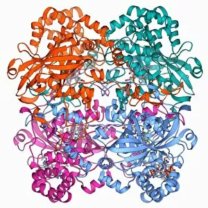
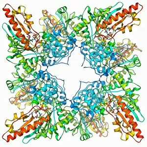
Citrate synthase, molecular model. This enzyme is involved in the first step of the citric acid (or Krebs) cycle, the process by which mitochondria convert glucose to energy

Background imageResearch Collection: Cobra venom molecule F006 / 9278

Cobra venom molecule F006 / 9278
Cobra venom. Molecular model of an alpha-cobratoxin (snake venom protein) from an Indochinese spitting cobra (Naja siamensis)

Background imageResearch Collection: DNA clamp complexed with DNA molecule F006 / 9274

DNA clamp complexed with DNA molecule F006 / 9274
DNA clamp complexed with DNA molecule. Molecular model showing a sliding DNA (deoxyribonucleic acid) clamp (beige) complexed with a molecule of DNA (red and blue)

Background imageResearch Collection: Synthetic triple helical peptide molecule F006 / 9275

Synthetic triple helical peptide molecule F006 / 9275
Synthetic triple helical peptide, molecular model

Background imageResearch Collection: mRNA capping enzyme molecule F006 / 9273

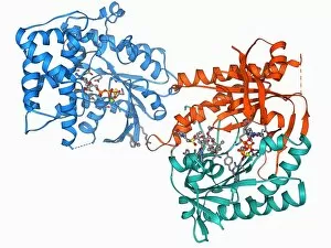
mRNA capping enzyme molecule F006 / 9273
mRNA capping enzyme. Molecular model of the mRNA capping enzyme mRNA guanylyltransferase complexed with a cap analogue

Background imageResearch Collection: Adenylyl cyclase enzyme molecule F006 / 9271

Adenylyl cyclase enzyme molecule F006 / 9271
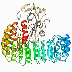
Adenylyl cyclase enzyme, molecular model. This enzyme catalyses the conversion of ATP (adenosine triphosphate) to cyclic AMP (cAMP) and pyrophosphate. cAMP regulates numerous cell functions

Background imageResearch Collection: Serotonin N-acetyl transferase molecule F006 / 9272

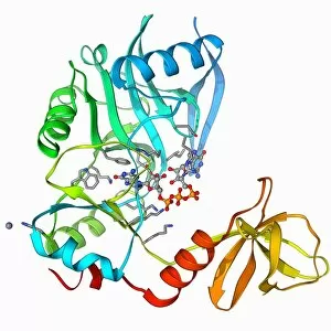
Serotonin N-acetyl transferase molecule F006 / 9272
Serotonin N-acetyl transferase. Molecular model of serotonin N-acetyl transferase complexed with coenzyme A-S-acetyltryptamine

Background imageResearch Collection: HGPRTase molecule F006 / 9270

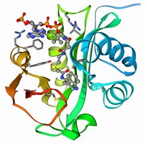
HGPRTase molecule F006 / 9270
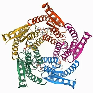
HGPRTase. Molecular model of hypoxanthine-guanine phosphoribosyltransferase (HGPRTase) from the malaria parasite Plasmodium falciparum. HGPRTase is involved in the purine salvage pathway


For sale as Licensed Images

Choose your image, Select your licence and Download the media

"Unveiling the Secrets: Exploring the Vast Realm of Research" From deciphering the valorous tales behind British Military medals to unraveling the enigmatic patterns in a Rorschach Inkblot Test, research takes us on an intellectual odyssey. It delves into realms beyond our comprehension, like scrutinizing the cosmic microwave background through MAP and decoding intricate histological diagrams of a mammalian retina. Research is not confined to laboratories; it extends to observing nature's wonders firsthand. Jane Goodall, with her unwavering dedication as a British conservationist and zoologist, captured alongside a chimpanzee, epitomizes how research bridges gaps between species. The quest for knowledge transcends boundaries of time and space. Erwin Schrodinger's groundbreaking discoveries in quantum mechanics remind us that research propels humanity forward by challenging conventional wisdom. In particle physics, proton collisions such as C014 / 1797 or witnessing Higgs boson events like C013 / 6892 within ATLAS detectors demonstrate how researchers strive to unlock mysteries at subatomic levels. Pioneers like Marie Curie exemplify relentless pursuit despite adversities faced by women in science. Her revolutionary work with radioactivity continues to inspire generations. It also embarks on daring expeditions akin to HMS Beagle - Darwin's ship that sailed uncharted waters - pushing boundaries of exploration and understanding. Similarly, Apollo 17 astronauts left indelible footprints on the Moon while expanding human horizons beyond Earth's confines. These captivating glimpses into diverse facets highlight its transformative power. It fuels curiosity, challenges assumptions, and empowers us with knowledge that shapes our world. Whether investigating microscopic particles or exploring vast ecosystems – research illuminates paths towards progress and inspires future generations to embark upon their own quests for discovery.