Retinal Collection (#2)

"Exploring the Intricacies of Retinal: Unveiling the Wonders of Vision" The retina, also known as the innermost layer of the eye

Background imageRetinal Collection: Cell death

Cell death. Computer-enhanced confocal light micrograph of cells in the retina of the eye undergoing programmed cell death (apoptosis)

Background imageRetinal Collection: Retina, light micrograph

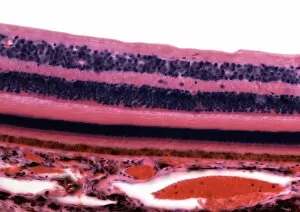
Retina, light micrograph
Retina. Light micrograph of a section through the retina showing its 10 layers. Magnification: x250 when printed at 10 centimetres wide

Background imageRetinal Collection: Cryptochrome and rhodopsin pigments

Cryptochrome and rhodopsin pigments. Rhodopsin is a pigment found in the rod photoreceptor cells in the retina of the human eye, where it absorbs light falling on the retina

Background imageRetinal Collection: Retina blood vessel, SEM

Retina blood vessel, SEM
Retina blood vessel. Coloured scanning electron micrograph (SEM) of a section through a blood vessel in the retina of an eye, showing the red blood cells (red)

Background imageRetinal Collection: Retina structure

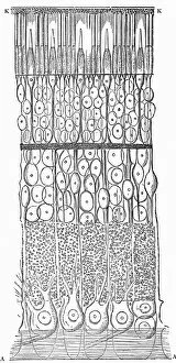
Retina structure
Retinal structure, artwork. Light falling on the retina passes from top to bottom. At bottom is the choroid layer (red), which lines the inside of the eye

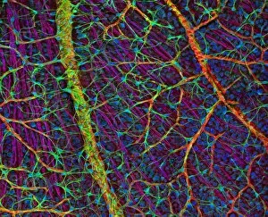
Background imageRetinal Collection: Retina blood vessels and nerve cells

Retina blood vessels and nerve cells. Light micrograph showing the blood vessels and nerve cells in the retina of an eye. The retina is the light-sensitive membrane that lines the back of the eyeball

Background imageRetinal Collection: Bacteriorhodopsin, diagram

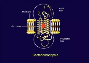
Bacteriorhodopsin, diagram
Bacteriorhodopsin. Diagram of the molecular structure of bacteriorhodopsin, a protein found in primitive micro-organisms known as Archaea

Background imageRetinal Collection: Bacteriorhodopsin, artwork

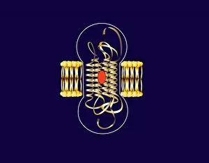
Bacteriorhodopsin, artwork
Bacteriorhodopsin. Artwork of the molecular structure of bacteriorhodopsin, a protein found in primitive micro-organisms known as Archaea

Background imageRetinal Collection: Human retina, 19th century artwork

Human retina, 19th century artwork. Artwork from the 1886 ninth edition of Moses and Geology (Samuel Kinns, London). This book was originally published in 1882

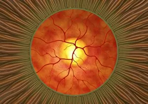
Background imageRetinal Collection: Retina in glaucoma, artwork

Retina in glaucoma, artwork
Retina in glaucoma, computer artwork. This view is looking through the pupil (green ring) at the front of the eye, to the retina (orange) at the back of the eye

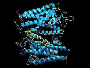
Background imageRetinal Collection: Sensory rhodopsin transducer molecule

Sensory rhodopsin transducer molecule. Computer model showing the molecular structure of sensory rhodopsin transducer (SRT)

Background imageRetinal Collection: Gecko retina, SEM

Gecko retina, SEM
Gecko retina. Coloured scanning electron micrograph (SEM) of an oblique freeze-fracture through the retina of a gecko (family Gekkonidae)

Background imageRetinal Collection: Vitamin A (retinal) molecule

Vitamin A (retinal) molecule
Vitamin A. Molecular model of the retinal form of vitamin A. Retinal is one of three forms of vitamin A; the other two are retinol and retinoic acid

Background imageRetinal Collection: Rhodopsin protein molecule

Rhodopsin protein molecule. Computer model of a molecule of the protein opsin that forms part of the rhodopsin complex. Rhodopsin is found in the rod photoreceptor cells in the retina of the human


For sale as Licensed Images

Choose your image, Select your licence and Download the media

"Exploring the Intricacies of Retinal: Unveiling the Wonders of Vision" The retina, also known as the innermost layer of the eye, plays a vital role in our visual perception. Composed of specialized cells called rod and cone cells, it acts as a gateway for light to be transformed into electrical signals that are then transmitted to the brain. Through scanning electron microscopy (SEM), we can observe the intricate structure of these rod and cone cells (C014 / 4866). These microscopic wonders are responsible for different aspects of vision - rods excel in low-light conditions while cones enable color perception. Another fascinating aspect captured by SEM is the network of blood vessels within the retina. These delicate vessels ensure proper nourishment and oxygen supply to this essential sensory tissue (Retina blood vessels, SEM). Zooming further into retinal exploration, transmission electron microscopy (TEM) reveals intriguing details such as Rift Valley fever virus particles present within retinal tissues. This highlights how viruses can impact ocular health on a microscopic level (Rift Valley fever virus, TEM). Delving deeper into molecular structures involved in vision, we encounter rhodopsin protein molecules. Rhodopsin is an integral component found in rod cells that enables them to detect light stimuli efficiently (Rhodopsin protein molecule). Its interaction with light triggers a cascade leading to signal transduction processes crucial for sight. Microscopic examination using advanced techniques like TEM allows us to witness mesmerizing images showcasing various components involved in visual processing. From bacteriorhodopsin proteins associated with photosynthesis-like reactions (Bacteriorhodopsin protein) to metarhodopsin molecules contributing to phototransduction pathways (F006 / 9709), each element unravels new insights about our remarkable sense of sight.