
Ribosome Collection (#2)
"Unraveling the Intricate World of Ribosomes: The Molecular Architects of Life" Delving into the microscopic realm


For sale as Licensed Images
Choose your image, Select your licence and Download the media
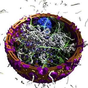
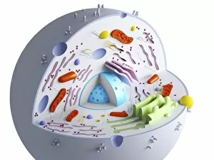
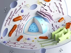
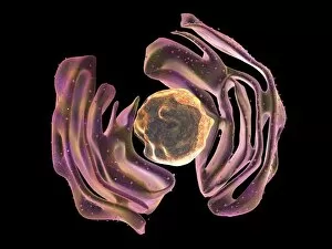
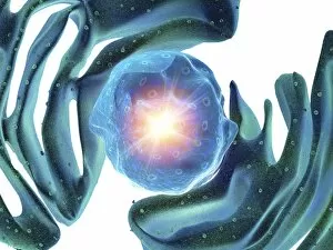
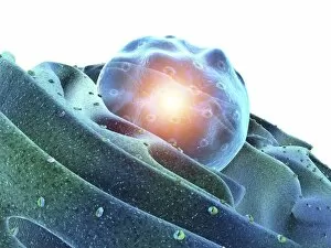
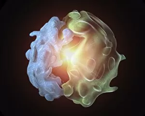
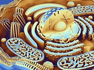
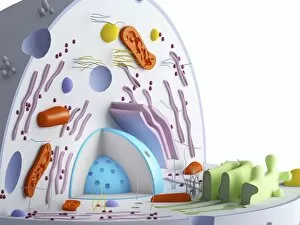
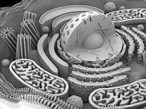
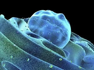
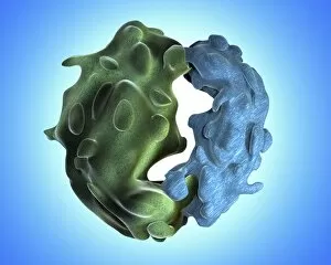
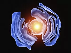
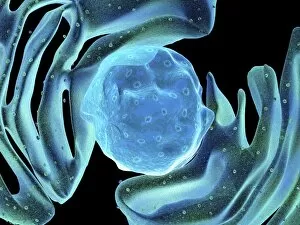
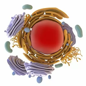
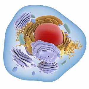
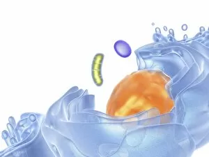
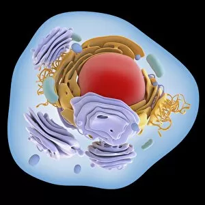
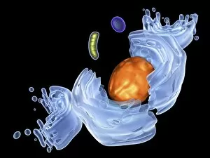
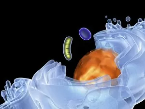
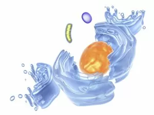
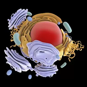
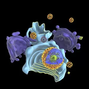
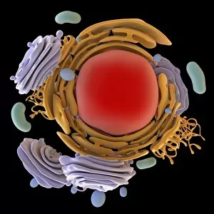
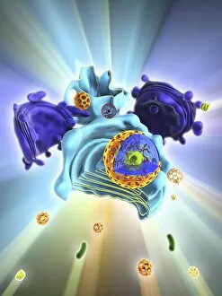
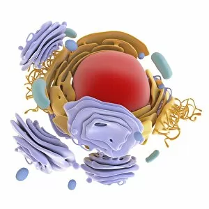
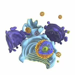
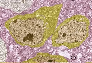
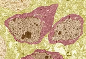
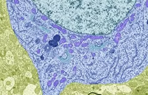
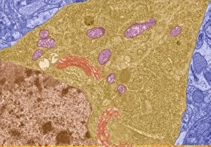
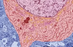
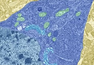
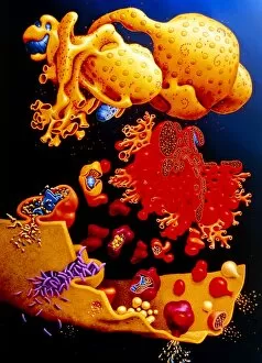
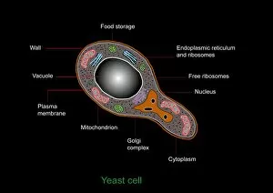
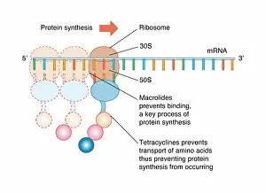
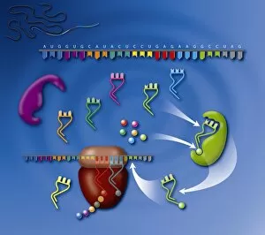
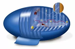
"Unraveling the Intricate World of Ribosomes: The Molecular Architects of Life" Delving into the microscopic realm, we encounter ribosomes - tiny but mighty cellular structures that orchestrate protein synthesis within living organisms. Found in various cell types, including nerve cells and animal cells, these intricate molecular machines play a pivotal role in sustaining life. Under the electron microscope's watchful eye, we witness ribosomes at work. In rough endoplasmic reticulum (ER), they dot its surface like diligent workers on an assembly line. This bustling ER serves as their workplace, ensuring efficient production of proteins crucial for cellular functions. Zooming closer to bacterial ribosomes reveals their simplicity yet remarkable efficiency. These streamlined versions lack the elaborate complexity seen in eukaryotic cells but are equally essential for bacterial survival and reproduction. In nerve cells captured by TEM imagery, ribosomes emerge as vital components responsible for synthesizing specialized proteins required for neuronal communication and function. Their presence highlights their indispensable role in maintaining our complex nervous system's integrity. Cross-section biomedical illustrations unveil the captivating process of protein synthesis guided by ribosomes. With precision akin to skilled architects constructing a building blueprint, these molecular maestros decode genetic information from DNA and assemble amino acids into functional proteins – fundamental building blocks of life itself. Within animal cell structures or human cell cross-sections lies another glimpse into this fascinating world. Nuclei surrounded by nucleoli house countless ribosomes diligently fulfilling their duty amidst a sea of organelles – all working harmoniously to sustain cellular vitality. Venturing beyond animals' borders leads us to plant cells where similar concepts prevail. Conceptual images showcase plant cell components such as nuclei, nucleoli, endoplasmic reticula intertwined with abundant ribosome activity – fueling growth and development while producing necessary proteins unique to plants' needs. As we explore further through microscopic lenses capturing animal or plant cells' intricacies, ribosomes emerge as silent heroes of cellular life.