
Ribs Collection (#7)
"Exploring the Intricacies of Ribs: From English Country Squire to Viking Ships and Beyond" Market Reports - English Country Squire carves the beef


For sale as Licensed Images
Choose your image, Select your licence and Download the media
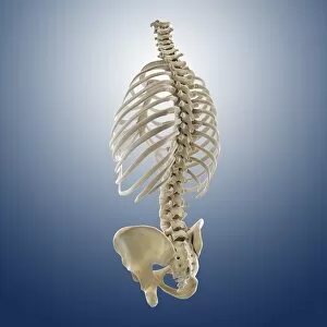
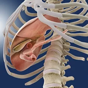
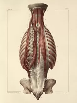
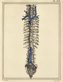
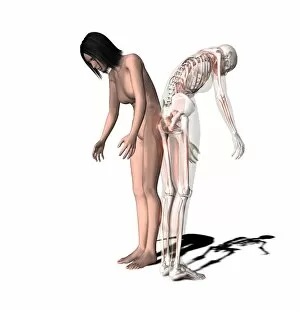
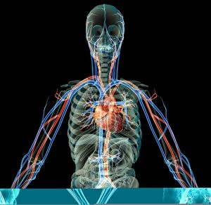
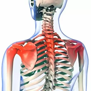
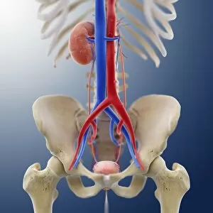
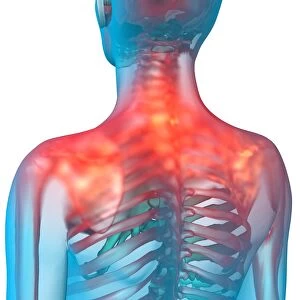
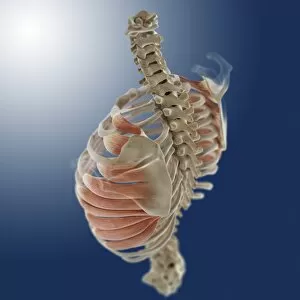
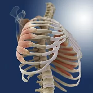
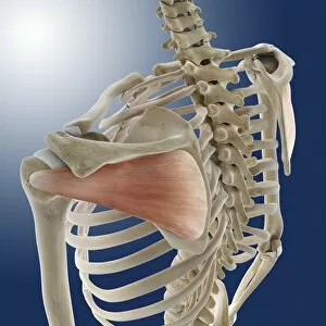
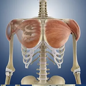
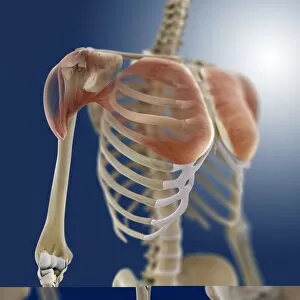
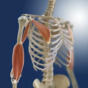
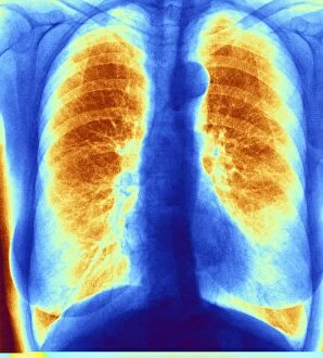
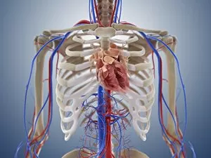
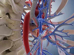
"Exploring the Intricacies of Ribs: From English Country Squire to Viking Ships and Beyond" Market Reports - English Country Squire carves the beef: Indulge in a succulent feast as the English Country Squire unveils his expertise, skillfully carving tender ribs that promise an unforgettable culinary experience. Cardiovascular system, historical artwork: Delve into the fascinating world of anatomy with historical artwork showcasing the intricate connection between our cardiovascular system and the protective cage of ribs. Nydam Viking Ship: Unearth ancient secrets as we voyage back to Viking times, where their iconic ships sailed across treacherous waters carrying warriors protected by sturdy ribcages. Upper body skeleton, computer artwork: Witness cutting-edge technology merging with anatomical precision as computer-generated imagery brings to life an upper body skeleton, highlighting the vital role played by ribs in safeguarding our organs. Neck and shoulder arteries, X-ray: Peer beneath the surface through an X-ray image revealing neck and shoulder arteries intertwined amidst a network of resilient ribs – a testament to our remarkable vascular system's resilience. Nude woman's torso, computer artwork: Admire artistry at its finest as computer-generated graphics present a stunning depiction of a nude woman's torso accentuating her graceful curves harmoniously protected by her delicate rib structure. Normal spine, X-ray: Unlock insights into spinal health with an illuminating X-ray capturing a normal spine flanked by robust ribs providing essential support for maintaining posture and protecting vital nerves. Tension pneumothorax, X-ray: Explore medical marvels through an X-ray image displaying tension pneumothorax – emphasizing how damaged or fractured they are lead to potentially life-threatening conditions requiring immediate attention. Skeletons, X-ray artwork: Marvel at artistic brilliance fused with scientific accuracy as mesmerizing x-ray artworks unveil skeletons adorned with intricately interwoven ribcages – reminding us of our shared skeletal framework regardless of our differences.