Ribs Collection (#9)

"Exploring the Intricacies of Ribs: From English Country Squire to Viking Ships and Beyond" Market Reports - English Country Squire carves the beef

Background imageRibs Collection: Diagnostic imaging

Diagnostic imaging. Conceptual computer artwork of a human figure with an exposed X-ray plate over his arm and ribcage. This may represent diagnostic medical imaging

Background imageRibs Collection: Healthy lung, X-ray

Healthy lung, X-ray
Healthy lung. Coloured X-ray of a healthy human lung. The spine runs vertically down far left. The ribs curve across the chest and protect the lungs. The upper arm bone (humerus) is at right

Background imageRibs Collection: Bronchioles

Bronchioles
Bronchiole network, composite image. The trachea (windpipe, not seen) branches into two bronchi, which enter the pair of lungs

Background imageRibs Collection: Lung, X-ray

Lung, X-ray
Lung. Coloured bronchography (X-ray) of a healthy human lung. A contrast medium has been added to show the network of airways (green) in the right lung

Background imageRibs Collection: Lungs and heart, X-ray

Lungs and heart, X-ray
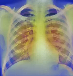
Lungs and heart, coloured chest X-ray. The lungs and heart are enclosed and protected by the ribs (horizontal bars) which meet at the breastbone, or sternum (down centre)

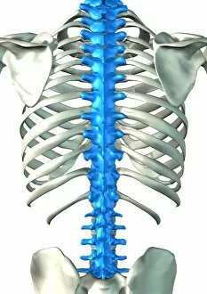
Background imageRibs Collection: Spine

Spine, computer artwork. Human skeleton with a blue spine. The spine, or backbone, supports the back and protects the spinal cord

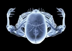
Background imageRibs Collection: Skeleton from above, X-ray artwork

Skeleton from above, X-ray artwork
Skeleton. Computer X-ray artwork of a human skeleton viewed from above

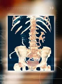
Background imageRibs Collection: Abdominal bones, 3-D CT scan

Abdominal bones, 3-D CT scan
Abdominal bones, coloured 3-D computer tomography (CT) scan. The lower part of the ribcage is in upper frame, with the pelvis in lower frame. They are connected by the spine, which runs down centre

Background imageRibs Collection: Spine, computer artwork

Spine, computer artwork
Spine. Computer artwork showing the bones of the spine, shoulder and pelvis on the human body. The spine, or backbone, supports the back and protects the spinal cord

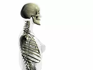
Background imageRibs Collection: Upper body skeleton

Upper body skeleton. Computer artwork of a glass- effect human female body in side view showing the skeletal structure of the head and torso

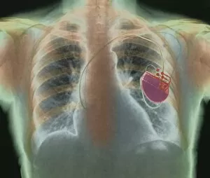
Background imageRibs Collection: Heart pacemaker, X-ray

Heart pacemaker, X-ray
Heart pacemaker. Coloured chest X-ray showing a surgically-implanted heart pacemaker (red, centre right). The pacemaker supplies electrical impulses along leads (running to lower centre)

Background imageRibs Collection: Sarcoidosis of the lungs, X-ray

Sarcoidosis of the lungs, X-ray
Sarcoidosis. Frontal coloured chest X-ray of the lungs of a patient with pulmonary sarcoidosis. Affected lung tissue shows up as red patches

Background imageRibs Collection: Lungs, X-ray

Lungs, X-ray

Background imageRibs Collection: Lungs, CT scans

Lungs, CT scans
Lungs. Sequence of six coloured axial CT (computed tomography) scans through the upper chest of a 52- year-old woman. The front of the chest is at top, and the right of the body is at left

Background imageRibs Collection: Heart, chest X-ray

Heart, chest X-ray

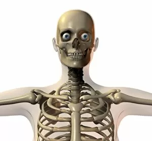
Background imageRibs Collection: Upper body bones

Upper body bones, computer artwork. The skull (top) consists of the cranium, which encloses the brain and the jaw (mandible)

Background imageRibs Collection: Ribcage

Ribcage and upper body, computer artwork. The spine, which consists of blocks of bone (vertebrae) separated by spongy intervertebral discs, runs down centre

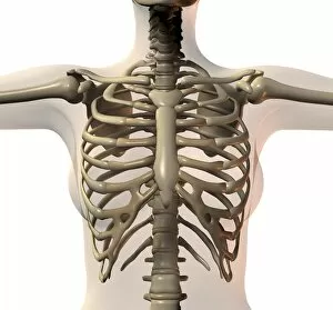
Background imageRibs Collection: Female ribcage

Female ribcage and upper body, computer artwork. The spine, which consists of blocks of bone (vertebrae) separated by spongy intervertebral discs, runs down centre

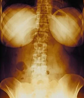
Background imageRibs Collection: Healthy lower back, X-ray

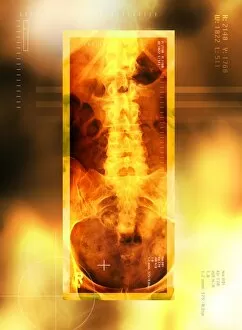
Healthy lower back, X-ray
Healthy lower back, coloured X-ray. The spine runs down centre. It is made up of vertebrae, block- shaped bones that support and protect the spinal cord (not seen)

Background imageRibs Collection: Torso nerves

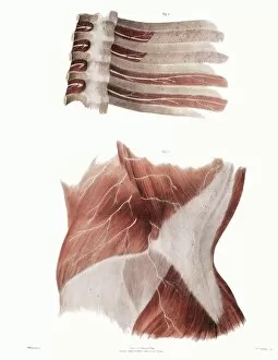
Torso nerves. Historical anatomical artwork of the nerves (white) of parts of a human torso. At lower frame, a side view of the abdomen (front of body at left) shows muscles (red)

Background imageRibs Collection: Spinal column

Spinal column. Historical artwork of the bones of the human spine and pelvis, seen from the front. The backbone (vertical)

Background imageRibs Collection: Spinal bones and ligaments

Spinal bones and ligaments. Historical anatomical artwork of thoracic (upper) spinal bones (yellow, vertebrae) and ligaments (white)

Background imageRibs Collection: Chronic bronchitis, X-ray

Chronic bronchitis, X-ray
Chronic bronchitis, coloured X-ray. This condition almost exclusively affects smokers. It is often associated with emphysema, which is known as COLD (chronic obstructive lung disease)

Background imageRibs Collection: Osteoporosis of spine, X-ray

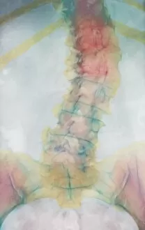
Osteoporosis of spine, X-ray
Osteoporosis. Coloured X-ray of a rear view of the lumbar (lower) spine of an elderly woman showing scoliosis (sideways curvature) of the spine caused by osteoporosis

Background imageRibs Collection: Bronchitis, X-ray

Bronchitis, X-ray
Bronchitis. Coloured bronchography (X-ray) of swollen bronchioles (orange) in the right lung. Bronchitis causes the network of airways in the lungs to become swollen and produce mucus

Background imageRibs Collection: Torso bones, computer artwork

Torso bones, computer artwork
Torso bones of a human skeleton seen from the front, computer artwork. The arm bones are raised to either side, with the base of the skull at top

Background imageRibs Collection: Female torso, X-ray

Female torso, X-ray
Female torso. Coloured frontal X-ray of a normal female torso, showing the bottom of the lungs (black, top), ribs radiating from upper centre, breasts (upper left and right)

Background imageRibs Collection: Radiologists examining an X-ray

Radiologists examining an X-ray
MODEL RELEASED. Radiologists studying an X-ray of a patients chest

Background imageRibs Collection: Human skeleton, computer artwork

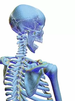
Human skeleton, computer artwork
Human skeleton. Computer artwork of the rear view of a human skeleton showing head and shoulders, superimposed with a wireframe

Background imageRibs Collection: Intestines, artwork

Intestines, artwork

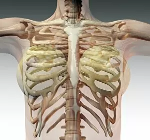
Background imageRibs Collection: Ribcage, computer artwork

Ribcage, computer artwork
Ribcage. Computer artwork of the pectoral girdle and ribcage as seen from the front

Background imageRibs Collection: Human internal organs, diagram

Human internal organs, diagram
Human internal organs. Diagram showing the human internal organs in the head and torso. In the head, the organs and cavities shown are: the brain (encephalon), the cerebellum (part of the brain)

Background imageRibs Collection: Human internal organs, artwork

Human internal organs, artwork
Human internal organs. Artwork showing the human internal organs in the head and torso. In the head, the organs and cavities shown are: the brain (encephalon), the cerebellum (part of the brain)


For sale as Licensed Images

Choose your image, Select your licence and Download the media

"Exploring the Intricacies of Ribs: From English Country Squire to Viking Ships and Beyond" Market Reports - English Country Squire carves the beef: Indulge in a succulent feast as the English Country Squire unveils his expertise, skillfully carving tender ribs that promise an unforgettable culinary experience. Cardiovascular system, historical artwork: Delve into the fascinating world of anatomy with historical artwork showcasing the intricate connection between our cardiovascular system and the protective cage of ribs. Nydam Viking Ship: Unearth ancient secrets as we voyage back to Viking times, where their iconic ships sailed across treacherous waters carrying warriors protected by sturdy ribcages. Upper body skeleton, computer artwork: Witness cutting-edge technology merging with anatomical precision as computer-generated imagery brings to life an upper body skeleton, highlighting the vital role played by ribs in safeguarding our organs. Neck and shoulder arteries, X-ray: Peer beneath the surface through an X-ray image revealing neck and shoulder arteries intertwined amidst a network of resilient ribs – a testament to our remarkable vascular system's resilience. Nude woman's torso, computer artwork: Admire artistry at its finest as computer-generated graphics present a stunning depiction of a nude woman's torso accentuating her graceful curves harmoniously protected by her delicate rib structure. Normal spine, X-ray: Unlock insights into spinal health with an illuminating X-ray capturing a normal spine flanked by robust ribs providing essential support for maintaining posture and protecting vital nerves. Tension pneumothorax, X-ray: Explore medical marvels through an X-ray image displaying tension pneumothorax – emphasizing how damaged or fractured they are lead to potentially life-threatening conditions requiring immediate attention. Skeletons, X-ray artwork: Marvel at artistic brilliance fused with scientific accuracy as mesmerizing x-ray artworks unveil skeletons adorned with intricately interwoven ribcages – reminding us of our shared skeletal framework regardless of our differences.